. 2020 Jan-Dec:29:963689720951549.
doi: 10.1177/0963689720951549.

Transplanted Antler Stem Cells Stimulated Regenerative Healing of Radiation-induced Cutaneous Wounds in Rats

Affiliations

Transplanted Antler Stem Cells Stimulated Regenerative Healing of Radiation-induced Cutaneous Wounds in Rats

Xiaoli Rong et al. Cell Transplant. 2020 Jan-Dec.

Abstract

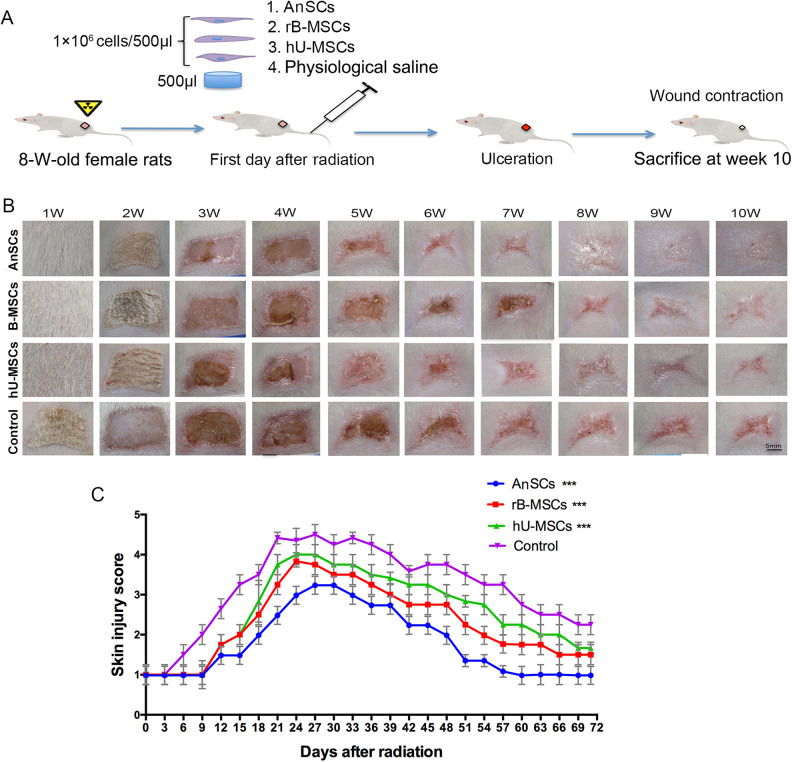
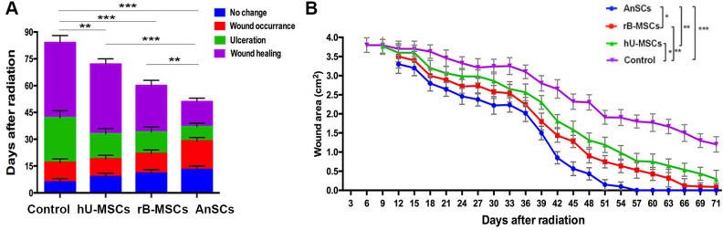
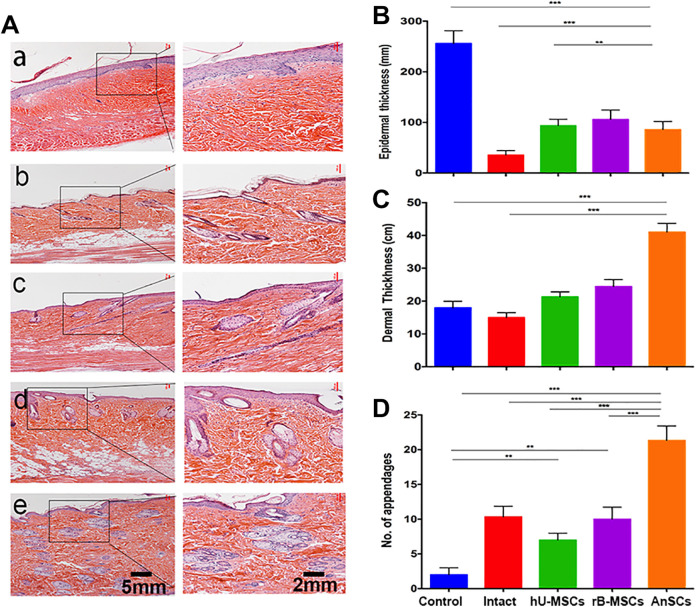
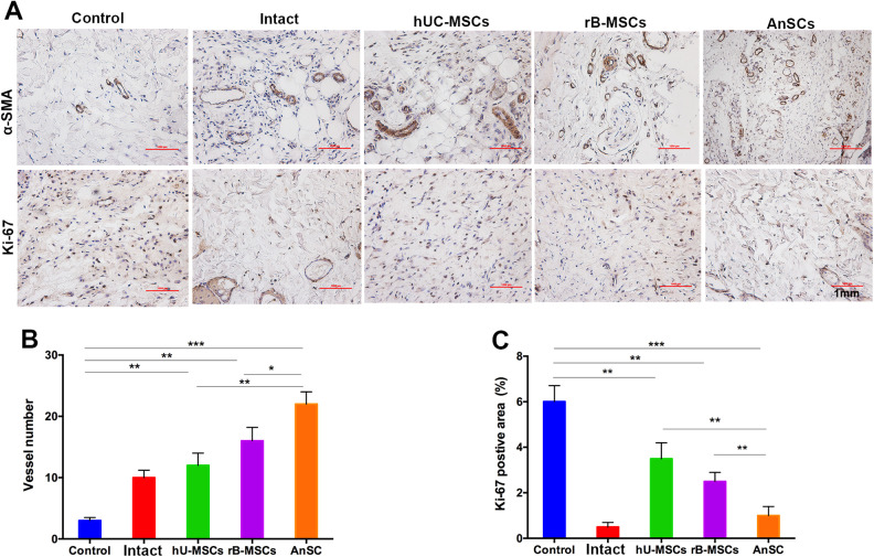
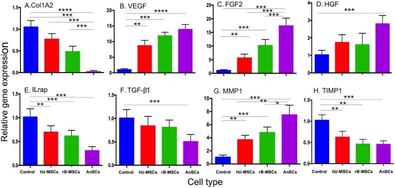
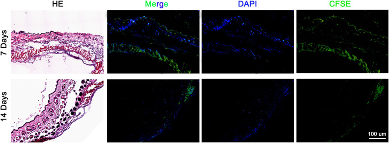
Radiation-induced cutaneous injury is the main side effect of radiotherapy. The injury is difficult to cure and the pathogenesis is complex. Mesenchymal stem cells (MSCs) serve as a promising candidate for cell-based therapy for the treatment of cutaneous wounds. The aim of the present study was to investigate whether antler stem cells (AnSCs) have better therapeutic effects on radiation-induced cutaneous injury than currently available ones. In this study, a rat model of cutaneous wound injury from Sr-90 radiation was used. AnSCs (1 × 10⁶/500 μl) were injected through the tail vein on the first day of irradiation. Our results showed that compared to the control group, AnSC-treated rats exhibited a delayed onset (14 days versus 7 days), shorter recovery time (51 days versus 84 days), faster healing rate (100% versus 70% on day 71), and higher healing quality with more cutaneous appendages regenerated (21:10:7/per given area compared to those of rat and human MSCs, respectively). More importantly, AnSCs promoted much higher quality of healing compared to other types of stem cells, with negligible scar formation. AnSC lineage tracing results showed that the injected-dye-stained AnSCs were substantially engrafted in the wound healing tissue, indicating that the therapeutic effects of AnSCs on wound healing at least partially through direct participation in the wound healing. Expression profiling of the wound-healing-related genes in the healing tissue of AnSC group more resembled a fetal wound healing. Revealing the mechanism underlying this higher quality of wound healing by using AnSC treatment would help to devise more effective cell-based therapeutics for radiation-induced wound healing in clinics.

Keywords: antler stem cells; cutaneous injury; radiation; wound healing.


Conflict of interest statement

Declaration of Conflicting Interests: The author(s) declare that they have no competing interests or any potential conflict of interest in any of the techniques or instruments mentioned in this article.

Figures

Figure 1.
Figure 1.
Comparison of proliferation rate among the three stem cell types. Number of the stem cells was counted following each subculture from one to eight passages. (A) The population doubling time was calculated based on cell counts (**P < 0.01, ***P < 0.001). (B) The data were shown as cumulative cell numbers at each passage. *P < 0.05, **P < 0.01 when compared to AnSC group, n = 3; mean ± SD. Each column represents the mean of the three stem cell populations with SD. AnSC: antler stem cell; SD: standard deviation.
Figure 2.
Figure 2.
Evaluation of the wound healing course. (A) Experimental procedures. (B) Gross morphological changes during wound occurring and healing. (C) Skin injuries were assessed using a semi-quantitative skin damage scores, with scores of 1.0 (no damage)–5.0 (severe damage). From day 15, all three stem-cell-treatment groups showed the lower scores than the control (***P < 0.001); n = 6; mean ± SD. SD: standard deviation.
Figure 3.
Figure 3.
Effects of stem cells on wound healing process and wound closure. (A) The accumulative column represents the time course of each cutaneous injury phase. (B) Changes in wound area during the course of wound closure. Multiple comparisons were made by one-way ANOVA. Data are reported as mean ± SD, n = 6. Dramatic wound area difference was found on day 39 as shown in top right corner (*P < 0.05, **P < 0.01, and ***P < 0.001). However, from days 27 to 54, the wound area was much smaller in the AnSC group than those of all other three groups (P < 0.05 to P < 0.001). From days 18 to 57, all the stem cell groups showed the significant lower wound areas than the control (P < 0.05 to P < 0.001); n = 6; mean ± SD. ANOVA: analysis of variance; AnSC: antler stem cell; SD: standard deviation.
Figure 4.
Figure 4.
Effects of the stem cells on histological structures of the healed skin tissue. (A) a–e: Control, Intact, hU-MSCs, rB-MSCs, and AnSCs, respectively. (B–D) Evaluation of epidermal and dermal thickness, and number of cutaneous appendages in the healed wound skin. Intact was taken from the same area as the other groups but without radiation. n = 6; mean ± SD (**P < 0.01, ***P < 0.001). AnSCs: antler stem cells; hU-MSCs: human umbilical cord mesenchymal stem cells; rB-MSCs: rat bone marrow mesenchymal stem cells; SD: standard deviation.
Figure 5.
Figure 5.
IHC evaluation of the wound healing quality. (A) Number of vessels/field (×100) in the healing tissue. (B) Positive staining area for Ki-67 analyzed using Image-Pro Plus. Note that AnSCs group had the highest number of vessels, but the least Ki-67 positive cells (on day 71 when wound healing reaching completion) compared to the rest of four groups. *P < 0.05, **P < 0.01, ***P < 0.001, bar = 1 mm; n = 6; mean ± SD. α-SMA: α-smooth muscle actin; AnSCs: antler stem cells; IHC: immunohistochemistry; SD: standard deviation.
Figure 6.
Figure 6.
Gene expression profiling of the healing skin tissue. Note that expression levels of the genes pro-wound healing in the MSC-treatment groups were all increased compared to the control group, whereas those of anti-wound healing in the MSC-treatment groups were all decreased compared to the control group. n = 6; mean ± SD (**P < 0.01, ***P < 0.001, and ****P < 0.0001). FGF2: fibroblast growth factor 2; HGF: hepatocyte growth factor; IL-1rap: interleukin-1 receptor accessory protein; MMP1: matrix metalloproteinase 1; MSC: mesenchymal stem cell; TGF-β1: transforming growth factor-β1; TIMP1: tissue inhibitor of metalloproteinase 1; VEGF: vascular endothelial growth factor.
Figure 7.
Figure 7.
AnSCs lineage tracing in the healing tissue on day 7 and day 14 after cell injection. Note that numerous dye-labeled AnSCs were detected in the dermal layer of healing tissue on day 7, although significantly reduced on day 14. AnSCs: antler stem cells.

References

    1. Singer AJ, Clark RA. Cutaneous wound healing. N Engl J Med. 1999;341(10):738–746. - PubMed
    1. Gourmelon P, Benderitter M, Bertho JM, Huet C, Gorin NC, De Revel P. European consensus on the medical management of acute radiation syndrome and analysis of the radiation accidents in Belgium and Senegal. Health Phys. 2010;98(6):825–832. - PubMed
    1. Ashcroft GS, Greenwell-Wild T, Horan MA, Wahl SM, Ferguson MW. Topical estrogen accelerates cutaneous wound healing in aged humans associated with an altered inflammatory response. Am J Pathol. 1999;155(4):1137–1146. - PMC - PubMed
    1. Cha J, Falanga V. Stem cells in cutaneous wound healing. Clin Dermatol. 2007;25(1):73–78. - PubMed
    1. Hocking AM. Mesenchymal stem cell therapy for cutaneous wounds. Adv Wound Care (New Rochelle). 2012;1(4):166–171. - PMC - PubMed

Publication types

MeSH terms