

Novel developmental bases for the evolution of hypobranchial muscles in vertebrates
- PMID: 32907560
- PMCID: PMC7488077
- DOI: 10.1186/s12915-020-00851-y
Novel developmental bases for the evolution of hypobranchial muscles in vertebrates
Abstract
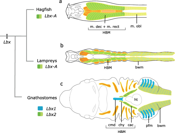
Background: Vertebrates are characterized by possession of hypobranchial muscles (HBMs). Cyclostomes, or modern jawless vertebrates, possess a rudimentary and superficial HBM lateral to the pharynx, whereas the HBM in jawed vertebrates is internalized and anteroposteriorly specified. Precursor cells of the HBM, marked by expression of Lbx1, originate from somites and undergo extensive migration before becoming innervated by the hypoglossal nerve. How the complex form of HBM arose in evolution is relevant to the establishment of the vertebrate body plan, but despite having long been assumed to be similar to that of limb muscles, modification of developmental mechanisms of HBM remains enigmatic.
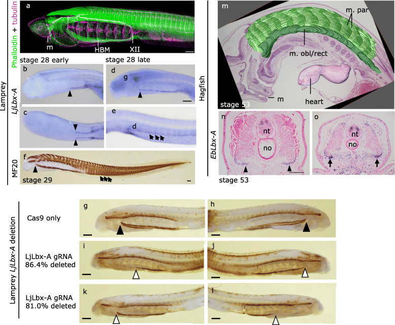
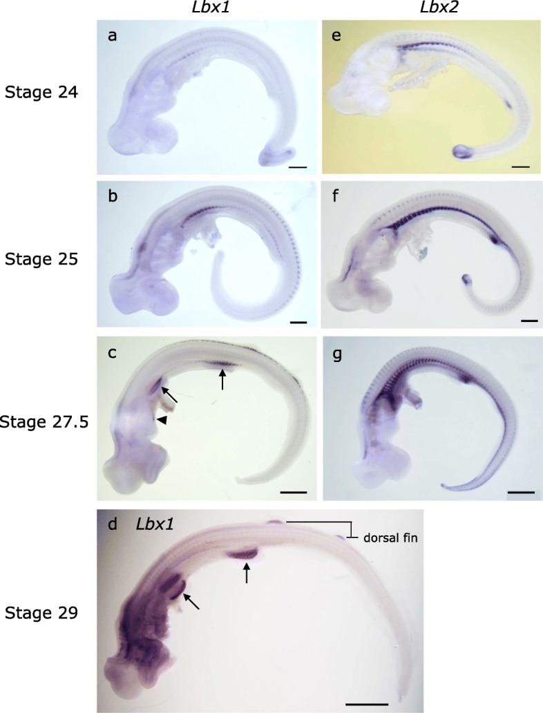
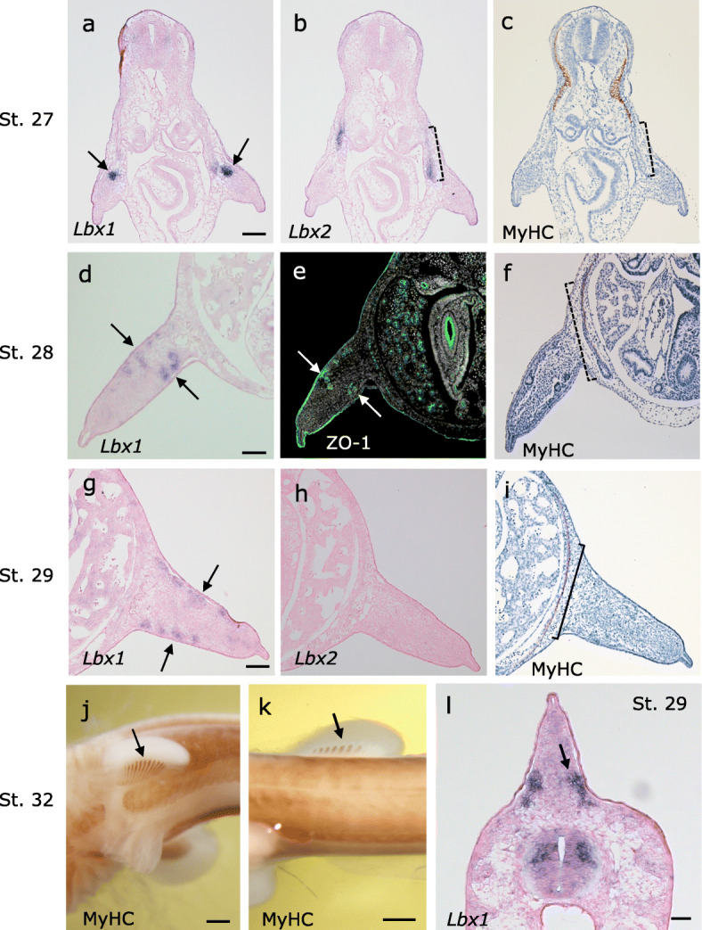
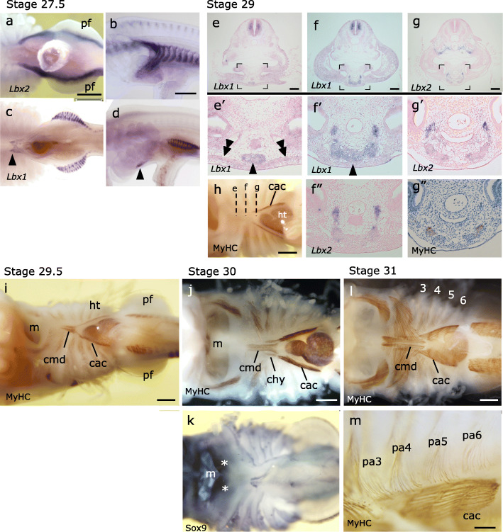
Results: Here we characterize the expression of Lbx genes in lamprey and hagfish (cyclostomes) and catshark (gnathostome; jawed vertebrates). We show that the expression patterns of the single cyclostome Lbx homologue, Lbx-A, do not resemble the somitic expression of mammalian Lbx1. Disruption of Lbx-A revealed that LjLbx-A is required for the formation of both HBM and body wall muscles, likely due to the insufficient extension of precursor cells rather than to hindered muscle differentiation. Both homologues of Lbx in the catshark were expressed in the somitic muscle primordia, unlike in amniotes. During catshark embryogenesis, Lbx2 is expressed in the caudal HBM as well as in the abdominal rectus muscle, similar to lamprey Lbx-A, whereas Lbx1 marks the rostral HBM and pectoral fin muscle.
Conclusions: We conclude that the vertebrate HBM primarily emerged as a specialized somatic muscle to cover the pharynx, and the anterior internalized HBM of the gnathostomes is likely a novelty added rostral to the cyclostome-like HBM, for which duplication and functionalization of Lbx genes would have been a prerequisite.
Keywords: Development; Evolution; Hagfish; Hypobranchial muscle; Lamprey; Lbx genes; Shark; Skeletal muscle; Vertebrates.
Conflict of interest statement
The authors declare that they have no competing interests.
Figures
Similar articles
-
Developmental Evolution of Hypaxial Muscles: Insights From Cyclostomes and Chondrichthyans.Front Cell Dev Biol. 2021 Sep 28;9:760366. doi: 10.3389/fcell.2021.760366. eCollection 2021. Front Cell Dev Biol. 2021. PMID: 34650989 Free PMC article. Review.
-
Expression and interaction of muscle-related genes in the lamprey imply the evolutionary scenario for vertebrate skeletal muscle, in association with the acquisition of the neck and fins.Dev Biol. 2011 Feb 1;350(1):217-27. doi: 10.1016/j.ydbio.2010.10.029. Epub 2010 Oct 28. Dev Biol. 2011. PMID: 21035440
-
Hagfish and lamprey Hox genes reveal conservation of temporal colinearity in vertebrates.Nat Ecol Evol. 2018 May;2(5):859-866. doi: 10.1038/s41559-018-0526-2. Epub 2018 Apr 2. Nat Ecol Evol. 2018. PMID: 29610468
-
Evolutionary crossroads in developmental biology: cyclostomes (lamprey and hagfish).Development. 2012 Jun;139(12):2091-9. doi: 10.1242/dev.074716. Development. 2012. PMID: 22619386 Review.
-
Development of hypobranchial muscles with special reference to the evolution of the vertebrate neck.Zoological Lett. 2018 Feb 18;4:5. doi: 10.1186/s40851-018-0087-x. eCollection 2018. Zoological Lett. 2018. PMID: 29468087 Free PMC article.
Cited by
-
Generation of knock-in lampreys by CRISPR-Cas9-mediated genome engineering.Sci Rep. 2021 Oct 6;11(1):19836. doi: 10.1038/s41598-021-99338-1. Sci Rep. 2021. PMID: 34615907 Free PMC article.
-
Thyroid and endostyle development in cyclostomes provides new insights into the evolutionary history of vertebrates.BMC Biol. 2022 Apr 1;20(1):76. doi: 10.1186/s12915-022-01282-7. BMC Biol. 2022. PMID: 35361194 Free PMC article.
-
Developmental Evolution of Hypaxial Muscles: Insights From Cyclostomes and Chondrichthyans.Front Cell Dev Biol. 2021 Sep 28;9:760366. doi: 10.3389/fcell.2021.760366. eCollection 2021. Front Cell Dev Biol. 2021. PMID: 34650989 Free PMC article. Review.
-
Spatial transcriptomics reveals a conserved segment polarity program that governs muscle patterning in Nematostella vectensis.bioRxiv [Preprint]. 2023 Jan 10:2023.01.09.523347. doi: 10.1101/2023.01.09.523347. bioRxiv. 2023. Update in: Curr Biol. 2023 Jul 10;33(13):2678-2689.e5. doi: 10.1016/j.cub.2023.05.044. PMID: 36711919 Free PMC article. Updated. Preprint.
References
-
- Mackenzie S, Walsh FS, Graham A. Migration of hypoglossal myoblast precursors. Dev Dyn. 1998;213(4):349–358. - PubMed
-
- Kuratani S. Spatial distribution of postotic crest cells defines the head/trunk interface of the vertebrate body: embryological interpretation of peripheral nerve morphology and evolution of the vertebrate head. Anat Embryol (Berl) 1997;195(1):1–13. - PubMed
-
- Christ B, Ordahl CP. Early stages of chick somite development. Anat Embryol (Berl) 1995;191(5):381–396. - PubMed
-
- Dietrich S, Schubert FR, Healy C, Sharpe PT, Lumsden A. Specification of the hypaxial musculature. Development. 1998;125(12):2235–2249. - PubMed
-
- Brohmann H, Jagla K, Birchmeier C. The role of Lbx1 in migration of muscle precursor cells. Development. 2000;127(2):437–445. - PubMed
Publication types
MeSH terms
Substances
Grants and funding
LinkOut - more resources
Full Text Sources
Research Materials
