

Plasma proteins facilitates placental transfer of polystyrene particles
- PMID: 32907583
- PMCID: PMC7487953
- DOI: 10.1186/s12951-020-00676-5
Plasma proteins facilitates placental transfer of polystyrene particles
Abstract
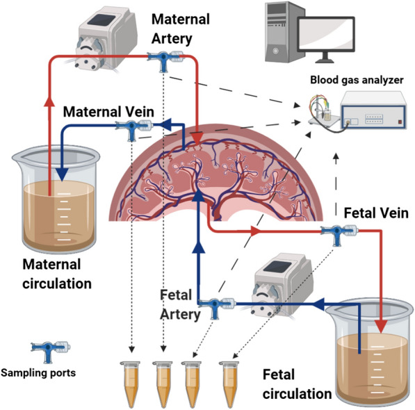
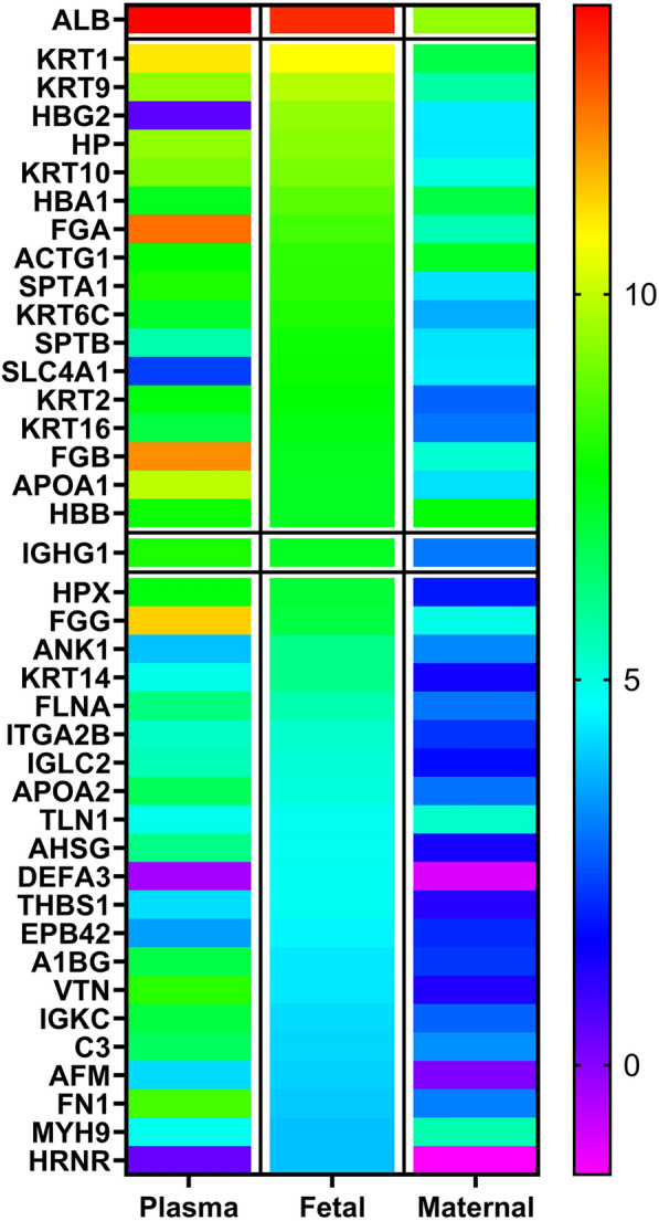
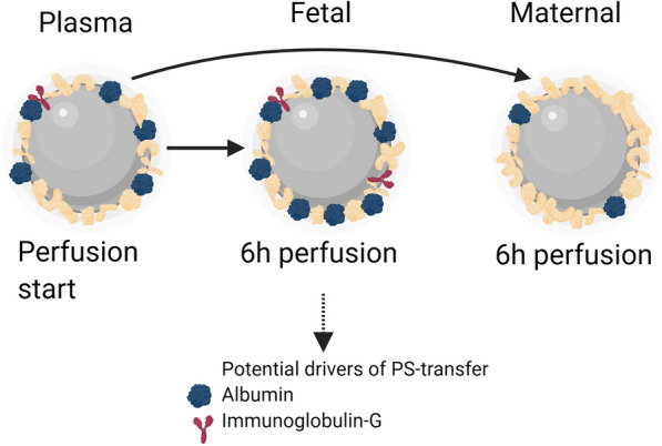
Background: Nanoparticles, which are exposed to biological fluids are rapidly interacting with proteins and other biomolecules forming a corona. In addition to dimension, charge and material the distinct protein corona influences the interplay of nanoparticles with tissue barriers. In this study we were focused on the impact of in situ formed human plasma protein corona on the transfer of 80 nm polystyrene nanoparticles (PS-particles) across the human placenta. To study materno-to fetal PS transfer we used the human ex vivo placental perfusion approach, which represents an intact and physiological tissue barrier. To analyze the protein corona of PS particles we performed shotgun proteomics of isolated nanoparticles before and after tissue exposure.
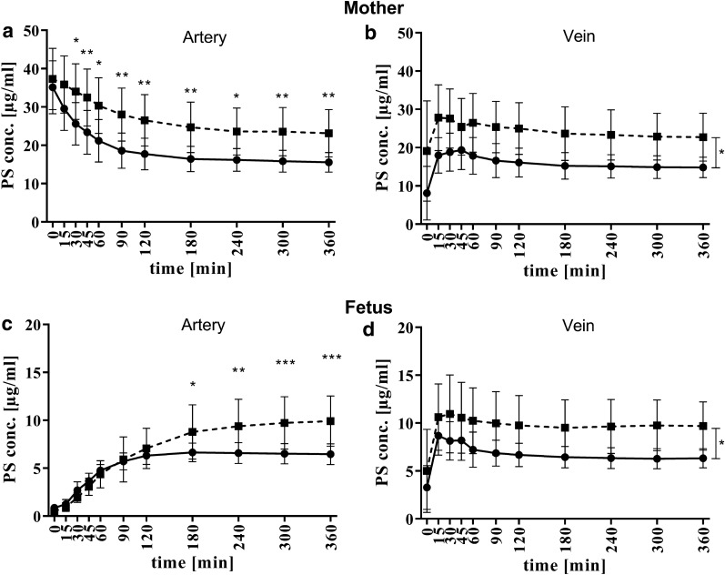
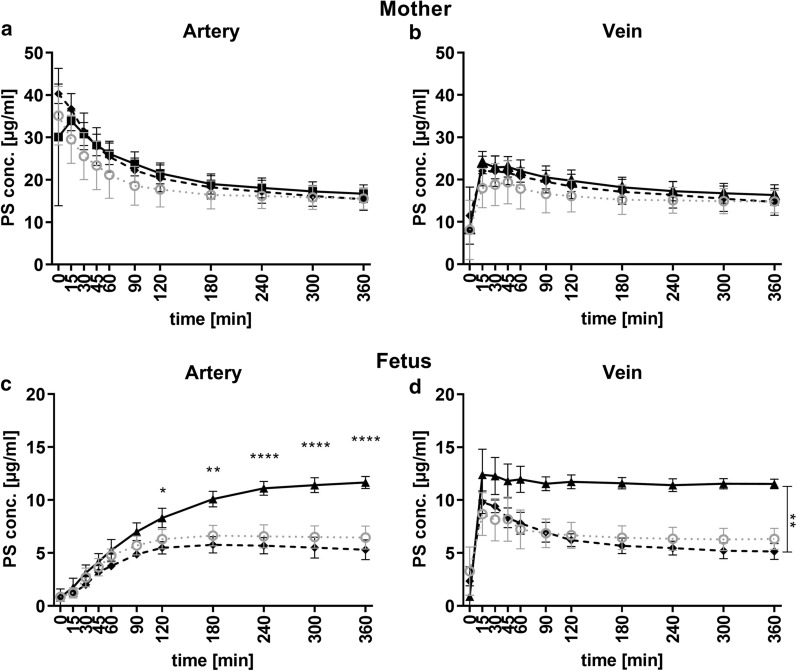
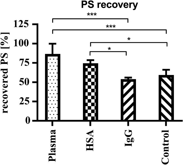
Results: Human plasma incubated with PS-particles of 80 nm and subsequent formed protein corona enhanced the transfer across the human placenta compared to PS-corona formed by bovine serum albumin and dextran which served as a control. Quantitative and qualitative changes of plasma proteins determined the changes in PS transfer across the barrier. Based on the analysis of the PS-proteome two candidate proteins, namely human albumin and immunoglobulin G were tested if these proteins may account for the enhanced PS-transfer across the placenta. Interestingly, the protein corona formed by human albumin significantly induced the transfer of PS-particles across the tissue compared to the formed IgG-corona.
Conclusion: In total we demonstrate the PS corona dynamically and significantly evolves upon crossing the human placenta. Thus, the initial composition of PS particles in the maternal circulation is not predictive for their transfer characteristics and performance once beyond the barrier of the placenta. The precise mechanism of these effects remains to be elucidated but highlights the importance of using well designed biological models when testing nanoparticles for biomedical applications.
Keywords: Biocorona; Dual ex vivo placental perfusion; Human placenta; Nanoparticle; Plasma proteins; Polystyrene; Transfer.
Conflict of interest statement
The authors declare that they have no competing interests.
Figures
References
-
- Tenzer S, Docter D, Kuharev J, Musyanovych A, Fetz V, Hecht R, et al. Rapid formation of plasma protein corona critically affects nanoparticle pathophysiology. Nat Nanotechnol. 2013;8:772–781. - PubMed
-
- Raesch SS, Tenzer S, Storck W, Rurainski A, Selzer D, Ruge CA, et al. Proteomic and lipidomic analysis of nanoparticle corona upon contact with lung surfactant reveals differences in protein, but not lipid composition. ACS Nano. 2015;9:11872–11885. - PubMed
-
- Ma X-X, Gao H, Zhang Y-X, Jia Y-Y, Li C, Zhou S-Y, et al. Construction and evaluation of BSA–CaP nanomaterials with enhanced transgene performance via biocorona-inspired caveolae-mediated endocytosis. Nanotechnology. 2018;29:085101. - PubMed
-
- Ke PC, Lin S, Parak WJ, Davis TP, Caruso F. A Decade of the Protein Corona. ACS Nano. 2017;11:11773–11776. - PubMed
MeSH terms
Substances
Grants and funding
LinkOut - more resources
Full Text Sources
Molecular Biology Databases
