

Chitosan-miRNA functionalized microporous titanium oxide surfaces via a layer-by-layer approach with a sustained release profile for enhanced osteogenic activity
- PMID: 32907598
- PMCID: PMC7487814
- DOI: 10.1186/s12951-020-00674-7
Chitosan-miRNA functionalized microporous titanium oxide surfaces via a layer-by-layer approach with a sustained release profile for enhanced osteogenic activity
Abstract
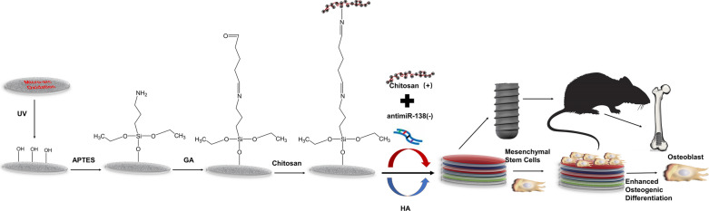
Background: The biofunctionalization of titanium implants for high osteogenic ability is a promising approach for the development of advanced implants to promote osseointegration, especially in compromised bone conditions. In this study, polyelectrolyte multilayers (PEMs) were fabricated using the layer-by-layer approach with a chitosan-miRNA (CS-miRNA) complex and sodium hyaluronate (HA) as the positively and negatively charged polyelectrolytes on microarc-oxidized (MAO) Ti surfaces via silane-glutaraldehyde coupling.
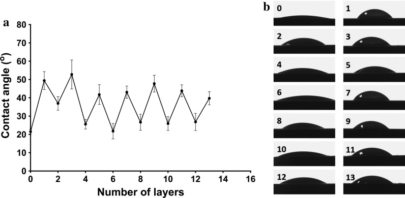
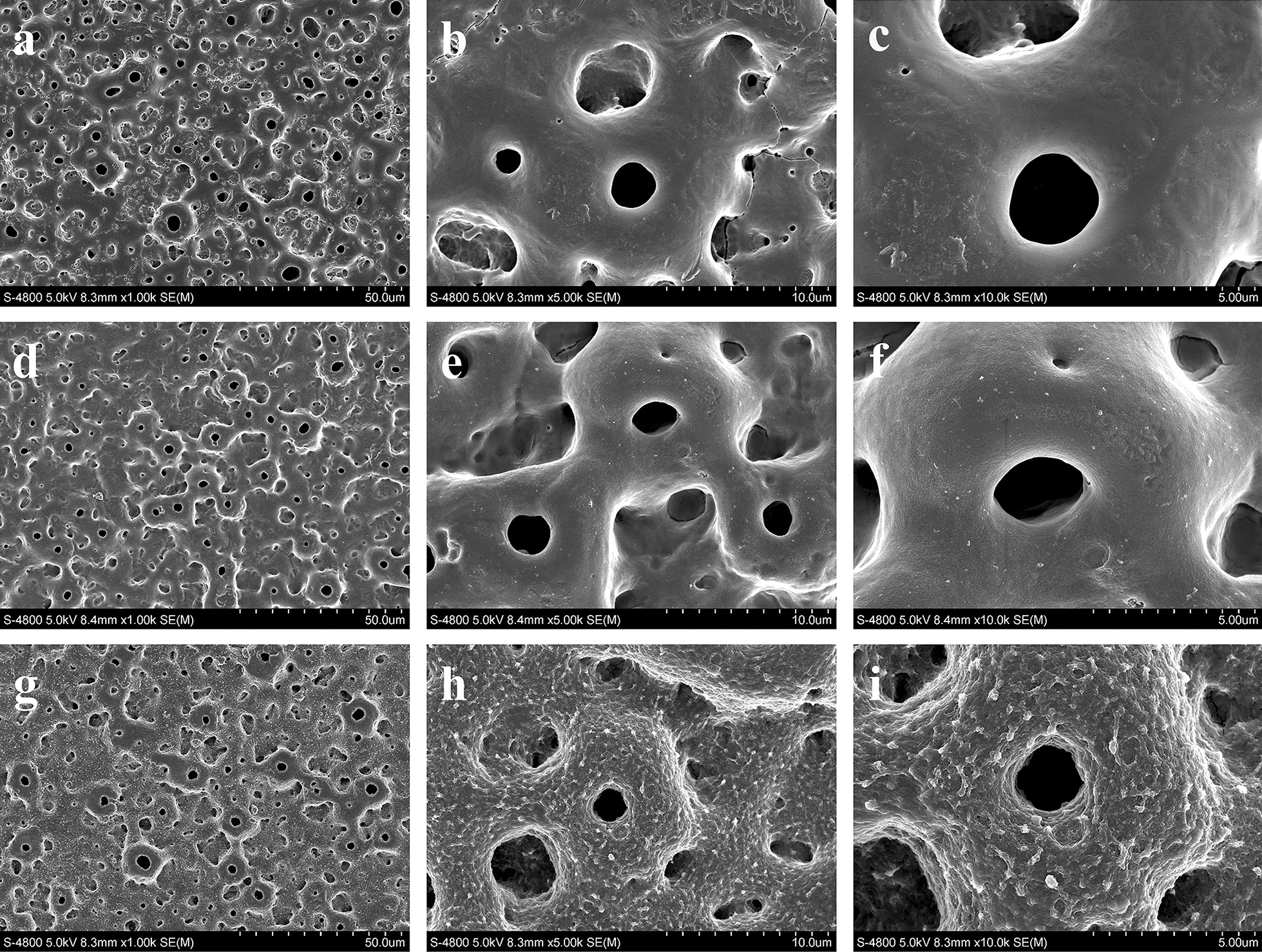
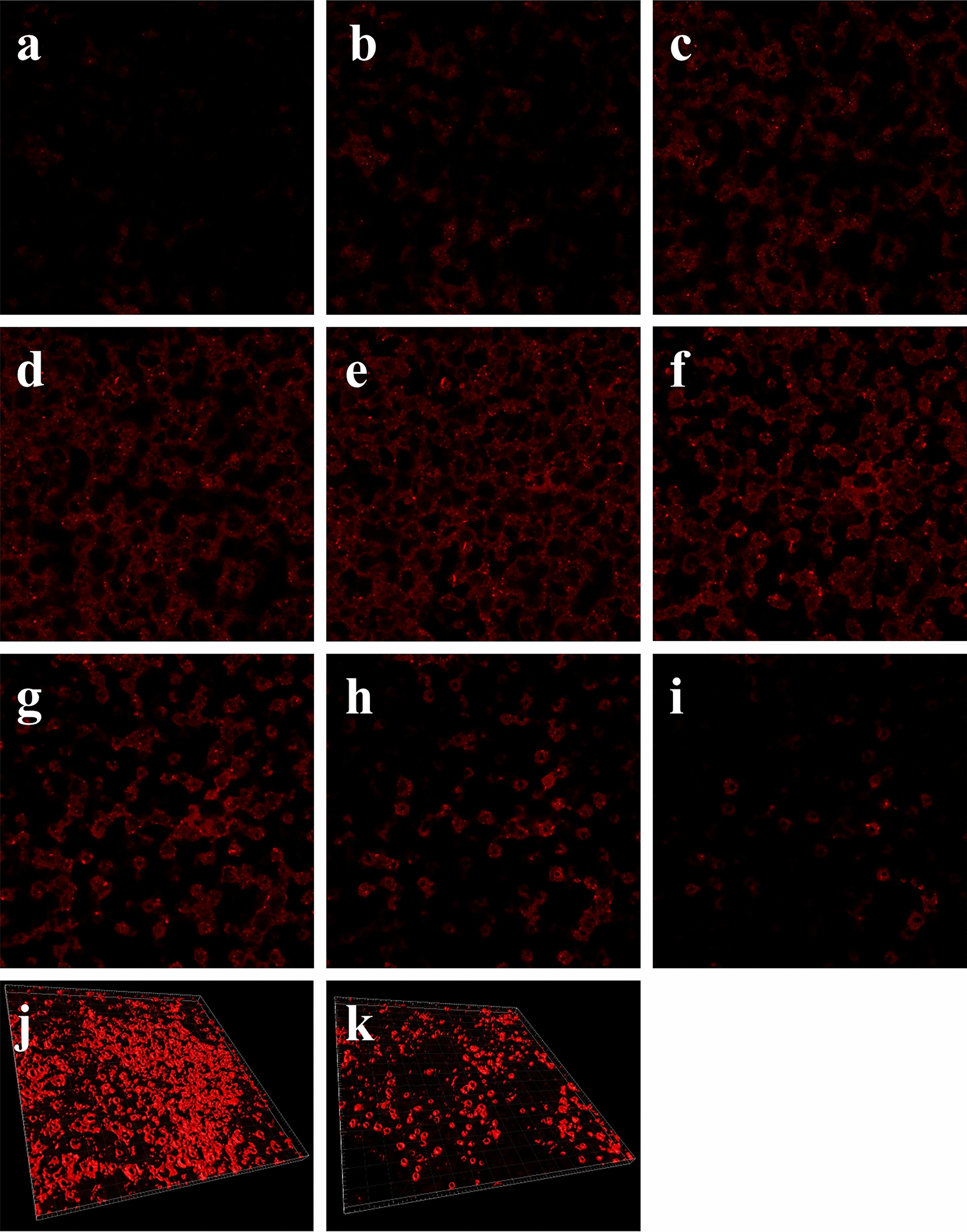
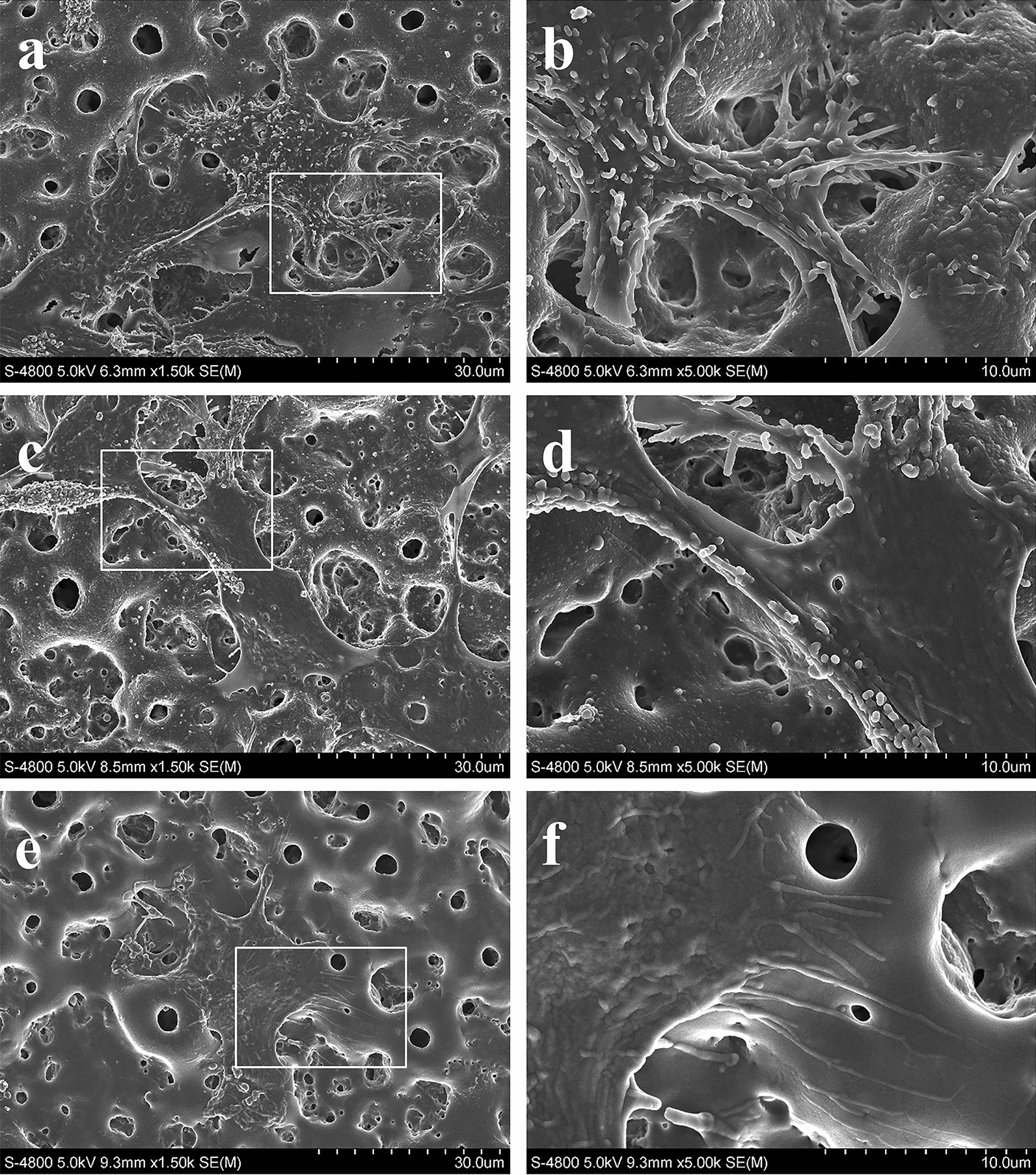
Methods: Dynamic contact angle and scanning electron microscopy measurements were conducted to monitor the layer accumulation. RiboGreen was used to quantify the miRNA loading and release profile in phosphate-buffered saline. The in vitro transfection efficiency and the cytotoxicity were investigated after seeding mesenchymal stem cells (MSCs) on the CS-antimiR-138/HA PEM-functionalized microporous Ti surface. The in vitro osteogenic differentiation of the MSCs and the in vivo osseointegration were also evaluated.
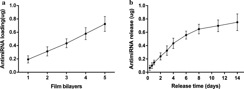
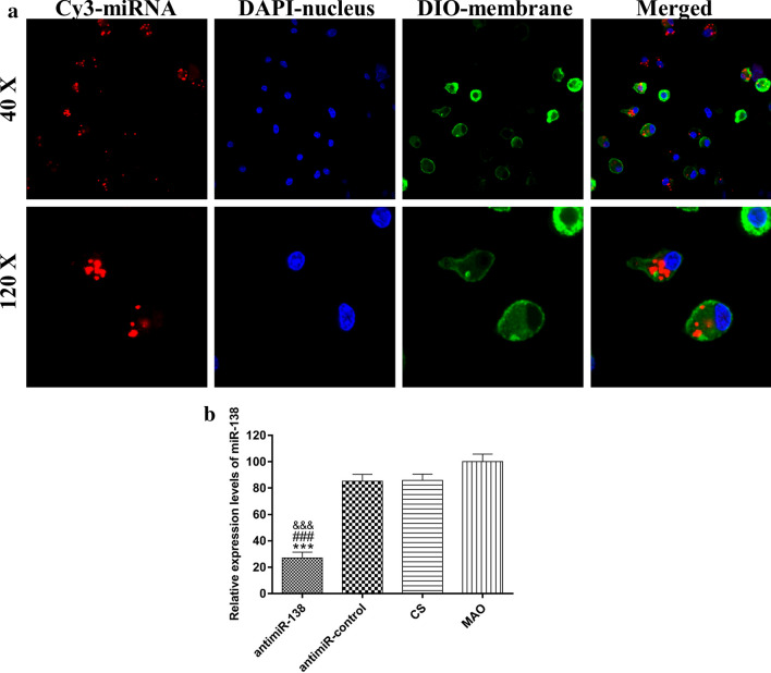
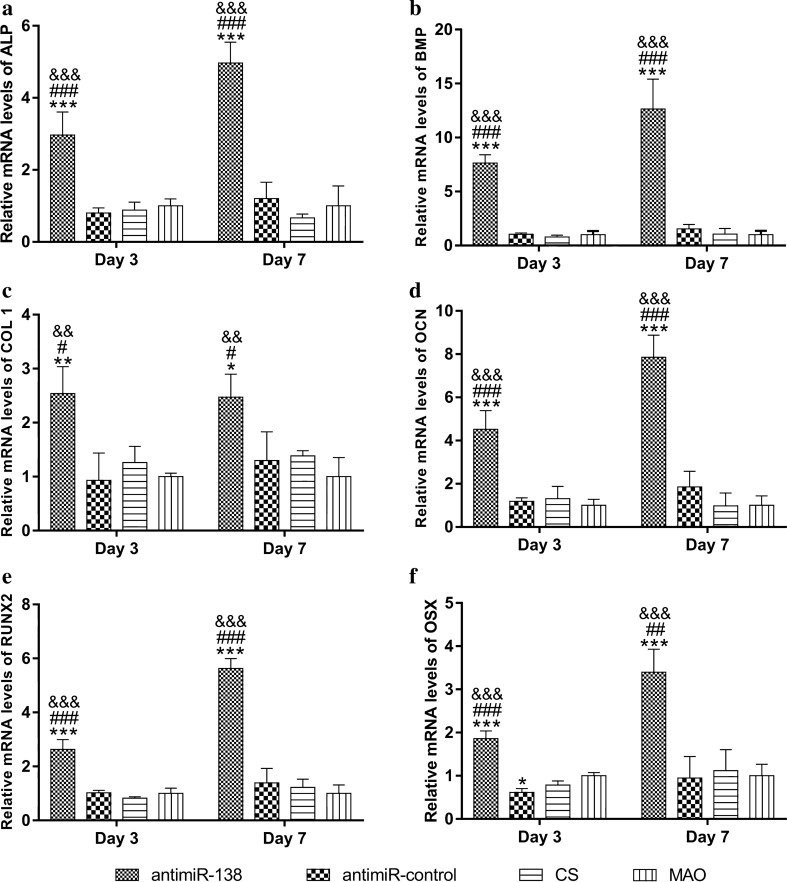
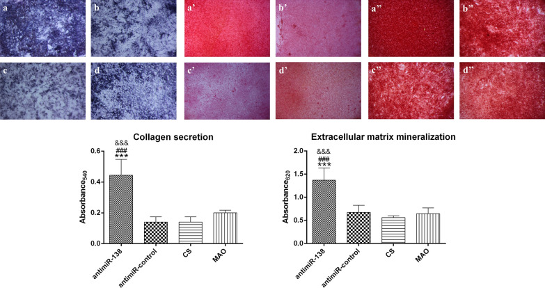
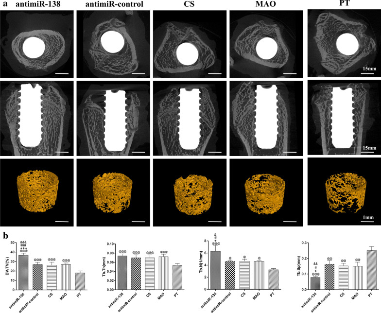
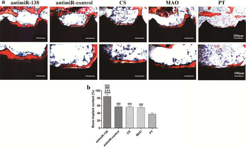
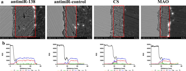
Results: The surface wettability alternately changed during the formation of PEMs. The CS-miRNA nanoparticles were distributed evenly across the MAO surface. The miRNA loading increased with increasing bilayer number. More importantly, a sustained miRNA release was obtained over a timeframe of approximately 2 weeks. In vitro transfection revealed that the CS-antimiR-138 nanoparticles were taken up efficiently by the cells and caused significant knockdown of miR-138 without showing significant cytotoxicity. The CS-antimiR-138/HA PEM surface enhanced the osteogenic differentiation of MSCs in terms of enhanced alkaline phosphatase, collagen production and extracellular matrix mineralization. Substantially enhanced in vivo osseointegration was observed in the rat model.
Conclusions: The findings demonstrated that the novel CS-antimiR-138/HA PEM-functionalized microporous Ti implant exhibited sustained release of CS-antimiR-138, and notably enhanced the in vitro osteogenic differentiation of MSCs and in vivo osseointegration. This novel miRNA-functionalized Ti implant may be used in the clinical setting to allow for more effective and robust osseointegration.
Keywords: Layer-by-layer; Mesenchymal stem cells; Microarc oxidation; Sustained release; Titanium implants; microRNAs.
Conflict of interest statement
The authors declare that they have no competing interests.
Figures
References
-
- Buser D, Sennerby L, De Bruyn H. Modern implant dentistry based on osseointegration: 50 years of progress, current trends and open questions. Periodontol. 2000;2017(73):7–21. - PubMed
-
- Civantos A, Martínez-Campos E, Ramos V, Elvira C, Gallardo A, Abarrategi A. Titanium coatings and surface modifications: toward clinically useful bioactive implants. ACS Biomater Sci Eng. 2017;3:1245–1261. - PubMed
-
- Buser D, Broggini N, Wieland M, Schenk RK, Denzer AJ, Cochran DL, Hoffmann B, Lussi A, Steinemann SG. Enhanced bone apposition to a chemically modified SLA titanium surface. J Dent Res. 2004;83:529–533. - PubMed
-
- Park JW, Jang JH, Lee CS, Hanawa T. Osteoconductivity of hydrophilic microstructured titanium implants with phosphate ion chemistry. Acta Biomater. 2009;5:2311–2321. - PubMed
-
- Iwata N, Nozaki K, Horiuchi N, Yamashita K, Tsutsumi Y, Miura H, Nagai A. Effects of controlled micro-/nanosurfaces on osteoblast proliferation. J Biomed Mater Res A. 2017;105:2589–2596. - PubMed
MeSH terms
Substances
Grants and funding
LinkOut - more resources
Full Text Sources
Research Materials
