

Preincubation with a low-dose hydrogen peroxide enhances anti-oxidative stress ability of BMSCs
- PMID: 32907609
- PMCID: PMC7487789
- DOI: 10.1186/s13018-020-01916-y
Preincubation with a low-dose hydrogen peroxide enhances anti-oxidative stress ability of BMSCs
Abstract
Objective: To investigate the effects of low-concentration hydrogen peroxide pretreatment on the anti-oxidative stress of the bone marrow mesenchymal stem cells (BMSCs).
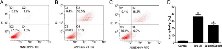
Methods: Rabbit BMSCs were isolated and cultured by density gradient centrifugation combined with the adherence method. Then, the third generation of well-grown BMSCs was continuously treated with 50-μM hydrogen peroxide (H2O2) for 8 h as the optimal pretreatment concentration and the BMSCs were continuously applied for 24 h with 500 μM H2O2, and the optimal damage concentration was determined as the oxidative stress cell model. The experiment was divided into three groups: control group, high-concentration H2O2 injury group (500 μM), and low-concentration H2O2 pretreatment group (50 μM + 500 μM). In each group, the DCFH-DA fluorescence probe was used to detect the reactive oxygen species (ROS). ELISA was used to detect the activity of superoxide dismutase (SOD) and catalase (CAT), and the TBA method was used to detect malondialdehyde (MDA). The mitochondrial membrane potential was detected by JC-1. The cell viability was detected by CCK-8 method, while flow cytometry and TUNEL/DAPI double staining were performed to detect cell apoptosis. Hence, the effect of H2O2 pretreatment on the anti-oxidative stress of BMSCs was investigated. One-way analysis of variance was performed using SPSS 19.0 statistical software, and P < 0.05 was considered statistically significant.
Results: A large number of typical BMSCs were obtained by density gradient centrifugation and adherent culture. The oxidative stress cell model was successfully established by 500-μM H2O2. Compared with the high-concentration H2O2 injury group, the low-concentration H2O2 pretreatment reduced the production of ROS [(62.33 ± 5.05), P < 0.05], SOD and CAT activities significantly increased (P < 0.05), and MDA levels significantly decreased (P < 0.05). The mitochondrial membrane potential fluorescence changes, the ratio of red/green fluorescence intensity of the high-concentration H2O2 injury group was less, and the ratio of the low-concentration H2O2 pretreatment group was significantly higher than that. The ratio of red/green increased by about 1.8 times (P < 0.05). The cell viability and survival rate of BMSCs were significantly increased in low-concentration H2O2 pretreatment group (P < 0.05), and the cell apoptosis rate was significantly decreased (P < 0.05).
Conclusion: Pretreatment with low-concentration H2O2 can enhance the anti-oxidative stress ability and reduce their apoptosis of BMSCs under oxidative stress.
Keywords: Bone marrow mesenchymal stem cells; Hydrogen peroxide, Apoptosis; Oxidative stress injury; Pretreatment.
Conflict of interest statement
The authors declare that they have no competing interests.
Figures


Similar articles
-
[Effects and mechanism of pyrroloquinoline quinine on mitochondrial function and cell survival of rat bone marrow mesenchymal stem cells under oxidative stress].Zhonghua Shao Shang Za Zhi. 2020 May 20;36(5):378-387. doi: 10.3760/cma.j.cn501120-20190806-00335. Zhonghua Shao Shang Za Zhi. 2020. PMID: 32456375 Chinese.
-
[Oxidative stress preconditioning alleviates oxidative stress-induced damage of bone marrow mesenchymal stem cells].Xi Bao Yu Fen Zi Mian Yi Xue Za Zhi. 2021 Feb;37(2):105-112. Xi Bao Yu Fen Zi Mian Yi Xue Za Zhi. 2021. PMID: 33504415 Chinese.
-
[Effects of nicotinamide mononucleotide adenylyl transferase 3 on mitochondrial function and anti-oxidative stress of rabbit bone marrow mesenchymal stem cells via regulating nicotinamide adenine dinucleotide levels].Zhongguo Xiu Fu Chong Jian Wai Ke Za Zhi. 2020 May 15;34(5):621-629. doi: 10.7507/1002-1892.201910037. Zhongguo Xiu Fu Chong Jian Wai Ke Za Zhi. 2020. PMID: 32410431 Free PMC article. Chinese.
-
Balanced Duality: H2O2-Based Therapy in Cancer and Its Protective Effects on Non-Malignant Tissues.Int J Mol Sci. 2024 Aug 15;25(16):8885. doi: 10.3390/ijms25168885. Int J Mol Sci. 2024. PMID: 39201571 Free PMC article. Review.
-
Anti-oxidant potential of plants and probiotic spp. in alleviating oxidative stress induced by H2O2.Biomed Pharmacother. 2023 Sep;165:115022. doi: 10.1016/j.biopha.2023.115022. Epub 2023 Jun 17. Biomed Pharmacother. 2023. PMID: 37336149 Review.
Cited by
-
Oxidative Stress Response in Adipose Tissue-Derived Mesenchymal Stem/Stromal Cells.Int J Mol Sci. 2022 Nov 3;23(21):13435. doi: 10.3390/ijms232113435. Int J Mol Sci. 2022. PMID: 36362223 Free PMC article.
-
Genistein and Procyanidin B2 Reduce Carcinogen-Induced Reactive Oxygen Species and DNA Damage through the Activation of Nrf2/ARE Cell Signaling in Bronchial Epithelial Cells In Vitro.Int J Mol Sci. 2023 Feb 12;24(4):3676. doi: 10.3390/ijms24043676. Int J Mol Sci. 2023. PMID: 36835090 Free PMC article.
-
Propofol Induces the Expression of Nrf2 and HO-1 in Echinococcus granulosus via the JNK and p38 Pathway In Vitro.Trop Med Infect Dis. 2023 Jun 3;8(6):306. doi: 10.3390/tropicalmed8060306. Trop Med Infect Dis. 2023. PMID: 37368724 Free PMC article.
-
Role of NF-κB signaling pathway in H2O2-induced oxidative stress of hiPSCs.In Vitro Cell Dev Biol Anim. 2024 Oct;60(9):1021-1033. doi: 10.1007/s11626-024-00943-x. Epub 2024 Aug 12. In Vitro Cell Dev Biol Anim. 2024. PMID: 39134871
-
Feline umbilical cord-derived mesenchymal stem cells: isolation, identification, and antioxidative stress role through NF-κB signaling pathway.Front Vet Sci. 2023 May 25;10:1203012. doi: 10.3389/fvets.2023.1203012. eCollection 2023. Front Vet Sci. 2023. PMID: 37303730 Free PMC article.
References
-
- Thanan R, Techasen A, Hou B, Jamnongkan W, Armartmuntree N, Yongvanit P, Murata M. Development and characterization of a hydrogen peroxide-resistant cholangiocyte cell line: a novel model of oxidative stress-related cholangiocarcinoma genesis. Biochem Biophys Res Commun. 2015;464:182–188. - PubMed
-
- Salazar-Noratto GE, Luo G, Denoeud C, et al. Understanding and leveraging cell metabolism to enhance mesenchymal stem cell transplantation survival in tissue engineering and regenerative medicine applications. Stem Cells. 2020;38(1):22–33. - PubMed
MeSH terms
Substances
LinkOut - more resources
Full Text Sources
Other Literature Sources
Miscellaneous
