

Construction of polycistronic baculovirus surface display vectors to express the PCV2 Cap(d41) protein and analysis of its immunogenicity in mice and swine
- PMID: 32907618
- PMCID: PMC7487469
- DOI: 10.1186/s13567-020-00836-3
Construction of polycistronic baculovirus surface display vectors to express the PCV2 Cap(d41) protein and analysis of its immunogenicity in mice and swine
Abstract
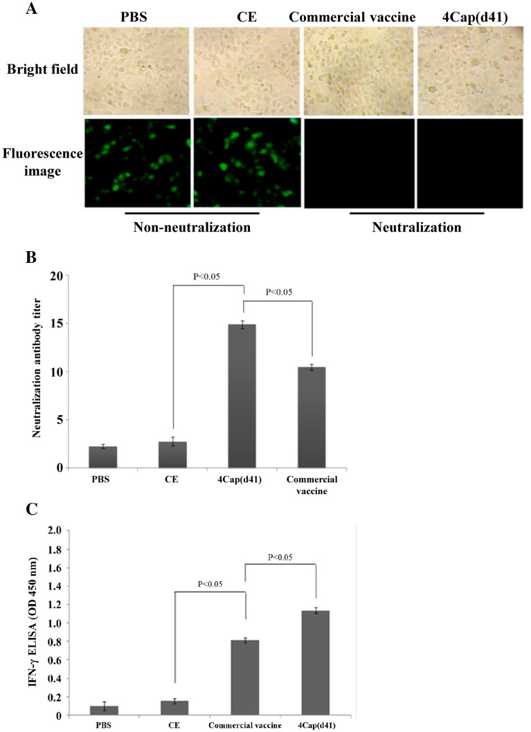
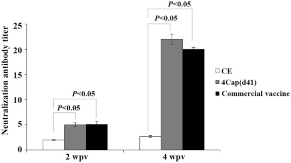
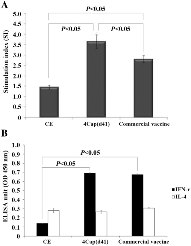
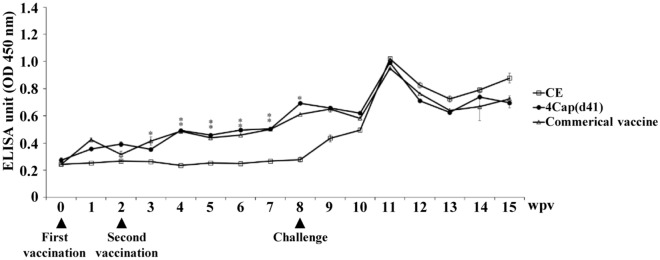
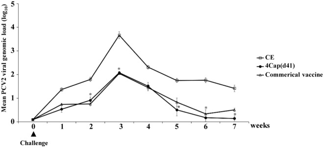
To increase expression levels of the PCV2 Cap(d41) protein, novel baculovirus surface display vectors with multiple expression cassettes were constructed to create recombinant baculoviruses BacSC-Cap(d41), BacDD-2Cap(d41), BacDD-3Cap(d41), and BacDD-4Cap(d41). Our results reveal that the recombinant baculovirus BacDD-4Cap(d41) was able to express the highest levels of Cap(d41) protein. Optimum conditions for expressing the PCV2 Cap(d41) protein were determined, and our results show that 107 of Sf-9 infected with the recombinant baculovirus BacDD-4Cap(d41) at an MOI of 5 for 3 days showed the highest level of protein expression. Mice immunized with the 4Cap(d41) vaccine which was prepared from the recombinant baculovirus-infected cells (107) elicited higher ELISA titers compared to the Cap (d41) vaccine. The 4Cap(d41) vaccine could elicit anti-PCV2 neutralizing antibodies and IFN-γ in mice, as confirmed by virus neutralization test and IFN-γ ELISA. Moreover, the swine lymphocyte proliferative responses indicated that the 4Cap(d41) vaccine was able to induce a clear cellular immune response. Flow cytometry analysis showed that the percentage of CD4+ T cells and CD4+/CD8+ ratio was increased significantly in SPF pigs immunized with the 4Cap(d41) vaccine. Importantly, the 4Cap(d41) vaccine induced an IFN-γ response, further confirming that its effect is through cellular immunity in SPF pigs. An in vivo challenge study revealed that the 4Cap(d41) and the commercial vaccine groups significantly reduce the viral load of vaccinated pigs as compared with the CE negative control group. Taken together, we have successfully developed a 4Cap(d41) vaccine that may be a potential subunit vaccine for preventing the disease associated with PCV2 infections.
Keywords: 4Cap(d41) vaccine; CD4+ T cells; Cap protein; IFN-γ; PCV2; baculovirus surface display vectors; cellular immune response; virus neutralization test.
Conflict of interest statement
The authors declare that they have no competing interests.
Figures
Similar articles
-
Baculovirus as a PRRSV and PCV2 bivalent vaccine vector: baculovirus virions displaying simultaneously GP5 glycoprotein of PRRSV and capsid protein of PCV2.J Virol Methods. 2012 Feb;179(2):359-66. doi: 10.1016/j.jviromet.2011.11.023. Epub 2011 Dec 6. J Virol Methods. 2012. PMID: 22172969
-
Oral vaccination with attenuated Salmonella enterica serovar Typhimurium expressing Cap protein of PCV2 and its immunogenicity in mouse and swine models.Vet Microbiol. 2012 Jun 15;157(3-4):294-303. doi: 10.1016/j.vetmic.2012.01.008. Epub 2012 Jan 17. Vet Microbiol. 2012. PMID: 22326539
-
Comparison of Immune Responses to the PCV2 Replicase-Capsid and Capsid Virus-Like Particle Vaccines in Mice.J Microbiol Biotechnol. 2019 Mar 28;29(3):482-488. doi: 10.4014/jmb.1809.09032. J Microbiol Biotechnol. 2019. PMID: 30609882
-
Establishment of a Rep' protein antibody detection method to distinguish natural infection with PCV2 from subunit vaccine immunization.J Med Microbiol. 2020 Sep;69(9):1183-1196. doi: 10.1099/jmm.0.001230. Epub 2020 Aug 19. J Med Microbiol. 2020. PMID: 32812860
-
Baculovirus Surface Display of Immunogenic Proteins for Vaccine Development.Viruses. 2018 May 31;10(6):298. doi: 10.3390/v10060298. Viruses. 2018. PMID: 29857561 Free PMC article. Review.
Cited by
-
The Magic Staff: A Comprehensive Overview of Baculovirus-Based Technologies Applied to Human and Animal Health.Viruses. 2022 Dec 28;15(1):80. doi: 10.3390/v15010080. Viruses. 2022. PMID: 36680120 Free PMC article. Review.
-
Baculovirus Display of Varicella-Zoster Virus Glycoprotein E Induces Robust Humoral and Cellular Immune Responses in Mice.Viruses. 2022 Aug 16;14(8):1785. doi: 10.3390/v14081785. Viruses. 2022. PMID: 36016407 Free PMC article.
-
Development of Polycistronic Baculovirus Surface Display Vectors to Simultaneously Express Viral Proteins of Porcine Reproductive and Respiratory Syndrome and Analysis of Their Immunogenicity in Swine.Vaccines (Basel). 2023 Oct 31;11(11):1666. doi: 10.3390/vaccines11111666. Vaccines (Basel). 2023. PMID: 38005998 Free PMC article.
-
Revisiting Porcine Circovirus Infection: Recent Insights and Its Significance in the Piggery Sector.Vaccines (Basel). 2023 Jul 31;11(8):1308. doi: 10.3390/vaccines11081308. Vaccines (Basel). 2023. PMID: 37631876 Free PMC article. Review.
-
Fusion Expression and Immune Effect of PCV2 Cap Protein Tandem Multiantigen Epitopes with CD154/GM-CSF.Vet Sci. 2021 Sep 29;8(10):211. doi: 10.3390/vetsci8100211. Vet Sci. 2021. PMID: 34679041 Free PMC article.
References
-
- Tischer I, Gelderblom H, Vettermann W, Koch MA. A very small porcine virus with circular single-stranded DNA. Nature. 1982;295:64–66. - PubMed
-
- Zhang HH, Hu WQ, Li JY, Liu TN, Zhou JY, Opriessnig T, Xiao CT. Novel circovirus species identified in farmed pigs designated as porcine circovirus 4, Hunan province, China. Transbound Emerg Dis. 2020;67(3):1057–1061. - PubMed
-
- Chae C. Postweaning multisystemic wasting syndrome: a review of aetiology, diagnosis and pathology. Vet J. 2004;168:41–49. - PubMed
-
- Mahe D, Blanchard P, Truong C, Arnauld C, Le Cann P, Cariolet R, Madec F, Albina E, Jestin A. Differential recognition of ORF2 protein from type 1 and type 2 procine circoviruses and identification of immunorelevant epitopes. J Gen Virol. 2000;81:1815–1824. - PubMed
MeSH terms
Substances
Grants and funding
LinkOut - more resources
Full Text Sources
Research Materials
Miscellaneous
