. 2020 Sep 22;117(38):23510-23518.
doi: 10.1073/pnas.2010000117. Epub 2020 Sep 9.

Dynamic structural order of a low-complexity domain facilitates assembly of intermediate filaments

Affiliations

Dynamic structural order of a low-complexity domain facilitates assembly of intermediate filaments

Vasiliy O Sysoev et al. Proc Natl Acad Sci U S A. .

Abstract

The coiled-coil domains of intermediate filament (IF) proteins are flanked by regions of low sequence complexity. Whereas IF coiled-coil domains assume dimeric and tetrameric conformations on their own, maturation of eight tetramers into cylindrical IFs is dependent on either "head" or "tail" domains of low sequence complexity. Here we confirm that the tail domain required for assembly of Drosophila Tm1-I/C IFs functions by forming labile cross-β interactions. These interactions are seen in polymers made from the tail domain alone, as well as in assembled IFs formed by the intact Tm1-I/C protein. The ability to visualize such interactions in situ within the context of a discrete cellular assembly lends support to the concept that equivalent interactions may be used in organizing other dynamic aspects of cell morphology.

Keywords: cross-beta polymerization; intermediate filaments; low-complexity proteins; solid-state NMR.


Conflict of interest statement

The authors declare no competing interest.

Figures
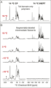
Fig. 1.
Fig. 1.
One-dimensional CP- and INEPT-based ss-NMR spectra of the Tm1-I/C tail domain. (A) CP (Left) and INEPT (Right) spectra of 13C/15N-labeled Tm1-I/C tail domain-only polymers recorded at 16 °C. (B) Four sets of spectra representing CP (Left) and INEPT (Right) spectra of segmentally 13C/15N-labeled Tm1-I/C protein assembled into mature intermediate filaments. Isotopic labeling was restricted to the Tm1-I/C tail domain by the use of intein chemistry as diagrammed in SI Appendix, Fig. S2A. The CP and INEPT spectra of fully assembled, segmentally labeled Tm1-I/C intermediate filaments were recorded at 16, 2, −12, and −19 °C as displayed from top to bottom. Asterisks in the top and bottom CP spectra indicate magic-angle spinning side bands.
Fig. 2.
Fig. 2.
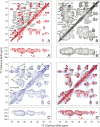
Two-dimensional CP-based ss-NMR spectra of the Tm1-I/C tail domain. (A and A′) 13C-13C CP-DARR spectrum with 50-ms mixing time of 13C/15N-labeled Tm1-I/C tail domain-only polymers recorded at 16 °C. Sequence-specific resonance assignments determined in this work are indicated by amino acid type and location within the full-length Tm1-I/C protein. (B and B′) The same spectrum of an ether-precipitated sample of the Tm1-I/C tail domain as analyzed in its polymeric form in A. Amino acid residues specified in the spectrum of ether precipitated protein were not assigned sequence-specifically. (C and C′) 13C-13C CP-DARR spectrum with 50-ms mixing time of segmentally 13C/15N-labeled Tm1-I/C protein that had been assembled into intermediate filaments recorded at −19 °C (SI Appendix, Fig. S2). Amino acid residues specified in the spectrum of segmentally labeled Tm1-I/C intermediate filaments were not assigned sequence-specifically. (D and D′) An overlay of the spectra shown in A and A′ and C and C′. The extensive overlap of signal intensity in the two samples indicates that the Tm1-I/C tail domain exists in highly similar structural states in the two assemblies. Sequence-specifically assigned amino acids from the data shown in A and A′ are indicated by small X’s in the overlay of D and D′. The spectra of the Tm1-I/C tail domain within assembled intermediate filaments (C and C′) overlap poorly or not at all with the spectra of ether-precipitated Tm1-I/C tail domain (B and B′). An overlay of the latter two spectra is shown in SI Appendix, Fig. S3.
Fig. 3.
Fig. 3.
Structural characterization of Tm1-I/C tail domain polymers. (A and A′) 2D CP-based NCACX spectrum of 13C/15N-labeled Tm1-I/C tail domain-only polymers measured at 16 °C. (B and B′) 2D 15N-13C zf-TEDOR spectrum of mixed (13C/14N)- and (12C/15N)-labeled Tm1-I/C tail domain-only polymers measured at 5 °C. (C) 2D 1H-13C INEPT-MAS spectra of 13C/15N-labeled Tm1-I/C tail domain-only polymers (red contours) overlapped with segmentally 13C/15N-labeled Tm1-I/C intermediate filaments (blue contours). Both spectra shown in C were recorded at 16 °C. (D) NMR chemical shift-based TALOS-N torsion angle predictions. Solid triangles and squares represent “strong” β-strand predictions, and open triangles and squares represent “generous” β-strand predictions determined according to published criteria (31).
Fig. 4.
Fig. 4.
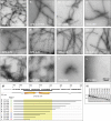
Functional assays of Tm1-I/C tail domain-only polymer formation for 10 C-terminal deletion mutants. Tm1-I/C tail domain variants missing 6, 11, 16, 21, 26, 31, 36, 41, 46, or 51 residues from the C terminus were expressed in bacterial cells, purified, and incubated under conditions suitable for tail domain polymerization. Samples were negatively stained with uranyl acetate and imaged by electron microscopy (Materials and Methods). (A and B) Intact tail domain polymers bearing an N-terminal His tag (A) or an N-terminal HA2 tag (B). (CE) Variants missing up to 16 amino acids formed polymers similar to the intact HA2-tagged Tm1-I/C tail domain. (FH) Variants missing between 21 and 31 amino acids formed distinctly twisted (T) polymers. (IK) Variants missing between 36 and 46 amino acids formed thin, spiky (S) polymers. (L) The variant missing 51 C-terminal amino acids did not form polymers. All electron micrographs were taken at the same magnification (Scale bar in L: 200 nm.) (M) Tm1-I/C tail deletion mutants relative to the amino acid sequence of the tail domain and the location of the cross-β core determined in this work (orange arrows and yellow highlight). (N) Coomassie brilliant blue-stained SDS polyacrylamide gel used to visualize the purified Tm1-I/C tail domain and 10 C-terminal deletion mutants. The numbers to the left of the SDS/PAGE gel indicate the migration positions of molecular weight markers at 15 and 10 kDa.
Fig. 5.
Fig. 5.
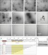
Functional assays of the ability of Tm1-I/C C-terminal deletion mutants to assemble into intermediate filaments. Deletion variants of the otherwise full-length protein missing 6, 11, 16, 21, 26, 31, 36, 41, 46, 51, or 69 C-terminal residues were expressed in bacterial cells, purified, and incubated under conditions allowing for the formation of intermediate filaments (Materials and Methods). (AD) Tm1-I/C variants missing up to 16 C-terminal residues of the protein formed intermediate filaments indistinguishable from those made from the intact protein. (EH) Variants missing between 21 and 36 C-terminal residues yielded aberrantly assembled filaments of a tangled or hairball-like morphology. (IL) Variants of the Tm1-I/C protein missing 41 or more C-terminal residues failed to form intermediate filaments. (M) Tm1-I/C tail domain deletion mutants relative to the amino acid sequence of the tail domain, the location of the C-terminal end of the coiled-coil domain (red), and the location of the cross-β core determined in this work (orange arrows and yellow highlight). (N) Coomassie brilliant blue-stained SDS polyacrylamide gel used to visualize purified samples of both intact (WT) Tm1-I/C and 11 C-terminal deletion mutants. The numbers to the left of the SDS/PAGE gel indicate the migration position of molecular weight markers at 100, 70, 55, and 40 kDa.

Similar articles

Cited by

References

    1. Trcek T., Lehmann R., Germ granules in Drosophila. Traffic 20, 650–660 (2019). - PMC - PubMed
    1. Ephrussi A., Lehmann R., Induction of germ cell formation by oskar. Nature 358, 387–392 (1992). - PubMed
    1. Gavis E. R., Lehmann R., Translational regulation of nanos by RNA localization. Nature 369, 315–318 (1994). - PubMed
    1. Bardsley A., McDonald K., Boswell R. E., Distribution of tudor protein in the Drosophila embryo suggests separation of functions based on site of localization. Development 119, 207–219 (1993). - PubMed
    1. Hay B., Jan L. Y., Jan Y. N., Localization of vasa, a component of Drosophila polar granules, in maternal-effect mutants that alter embryonic anteroposterior polarity. Development 109, 425–433 (1990). - PubMed

Publication types

MeSH terms

Substances