Continued Evolution of H5Nx Avian Influenza Viruses in Bangladeshi Live Poultry Markets: Pathogenic Potential in Poultry and Mammalian Models
- PMID: 32907981
- PMCID: PMC7654280
- DOI: 10.1128/JVI.01141-20
Continued Evolution of H5Nx Avian Influenza Viruses in Bangladeshi Live Poultry Markets: Pathogenic Potential in Poultry and Mammalian Models
Abstract
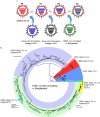
The genesis of novel influenza viruses through reassortment poses a continuing risk to public health. This is of particular concern in Bangladesh, where highly pathogenic avian influenza viruses of the A(H5N1) subtype are endemic and cocirculate with other influenza viruses. Active surveillance of avian influenza viruses in Bangladeshi live poultry markets detected three A(H5) genotypes, designated H5N1-R1, H5N1-R2, and H5N2-R3, that arose from reassortment of A(H5N1) clade 2.3.2.1a viruses. The H5N1-R1 and H5N1-R2 viruses contained HA, NA, and M genes from the A(H5N1) clade 2.3.2.1a viruses and PB2, PB1, PA, NP, and NS genes from other Eurasian influenza viruses. H5N2-R3 viruses contained the HA gene from circulating A(H5N1) clade 2.3.2.1a viruses, NA and M genes from concurrently circulating A(H9N2) influenza viruses, and PB2, PB1, PA, NP, and NS genes from other Eurasian influenza viruses. Representative viruses of all three genotypes and a parental clade 2.3.2.1a strain (H5N1-R0) infected and replicated in mice without prior adaptation; the H5N2-R3 virus replicated to the highest titers in the lung. All viruses efficiently infected and killed chickens. All viruses replicated in inoculated ferrets, but no airborne transmission was detected, and only H5N2-R3 showed limited direct-contact transmission. Our findings demonstrate that although the A(H5N1) viruses circulating in Bangladesh have the capacity to infect and replicate in mammals, they show very limited capacity for transmission. However, reassortment does generate viruses of distinct phenotypes.IMPORTANCE Highly pathogenic avian influenza A(H5N1) viruses have circulated continuously in Bangladesh since 2007, and active surveillance has detected viral evolution driven by mutation and reassortment. Recently, three genetically distinct A(H5N1) reassortant viruses were detected in live poultry markets in Bangladesh. Currently, we cannot assign pandemic risk by only sequencing viruses; it must be conducted empirically. We found that the H5Nx highly pathogenic avian influenza viruses exhibited high virulence in mice and chickens, and one virus had limited capacity to transmit between ferrets, a property considered consistent with a higher zoonotic risk.
Keywords: Bangladesh; influenza; influenza viruses; live poultry markets.
Copyright © 2020 American Society for Microbiology.
Figures


Similar articles
-
The Continuing Evolution of H5N1 and H9N2 Influenza Viruses in Bangladesh Between 2013 and 2014.Avian Dis. 2016 May;60(1 Suppl):108-17. doi: 10.1637/11136-050815-Reg. Avian Dis. 2016. PMID: 27309046 Free PMC article.
-
Continuing evolution of highly pathogenic H5N1 viruses in Bangladeshi live poultry markets.Emerg Microbes Infect. 2019;8(1):650-661. doi: 10.1080/22221751.2019.1605845. Emerg Microbes Infect. 2019. PMID: 31014196 Free PMC article.
-
Reassortment of newly emergent clade 2.3.4.4b A(H5N1) highly pathogenic avian influenza A viruses in Bangladesh.Emerg Microbes Infect. 2025 Dec;14(1):2432351. doi: 10.1080/22221751.2024.2432351. Epub 2024 Dec 9. Emerg Microbes Infect. 2025. PMID: 39584394 Free PMC article.
-
Review analysis and impact of co-circulating H5N1 and H9N2 avian influenza viruses in Bangladesh.Epidemiol Infect. 2018 Jul;146(10):1259-1266. doi: 10.1017/S0950268818001292. Epub 2018 May 21. Epidemiol Infect. 2018. PMID: 29781424 Free PMC article.
-
The genetics of highly pathogenic avian influenza viruses of subtype H5 in Germany, 2006-2020.Transbound Emerg Dis. 2021 May;68(3):1136-1150. doi: 10.1111/tbed.13843. Epub 2020 Sep 29. Transbound Emerg Dis. 2021. PMID: 32964686 Review.
Cited by
-
Semi-Scavenging Poultry as Carriers of Avian Influenza Genes.Life (Basel). 2022 Feb 21;12(2):320. doi: 10.3390/life12020320. Life (Basel). 2022. PMID: 35207607 Free PMC article.
-
Identification & genetic & virological characterisation of a human case of avian influenza A (H9N2) virus from Eastern India.Indian J Med Res. 2025 Mar;161(3):257-266. doi: 10.25259/IJMR_1376_2024. Indian J Med Res. 2025. PMID: 40347502 Free PMC article.
-
H9N2 avian influenza virus dispersal along Bangladeshi poultry trading networks.Virus Evol. 2023 Feb 25;9(1):vead014. doi: 10.1093/ve/vead014. eCollection 2023. Virus Evol. 2023. PMID: 36968264 Free PMC article.
-
Development of avian influenza A(H5) virus datasets for Nextclade enables rapid and accurate clade assignment.bioRxiv [Preprint]. 2025 Feb 3:2025.01.07.631789. doi: 10.1101/2025.01.07.631789. bioRxiv. 2025. PMID: 39829835 Free PMC article. Preprint.
-
The H5N6 Virus Containing Internal Genes From H9N2 Exhibits Enhanced Pathogenicity and Transmissibility.Transbound Emerg Dis. 2025 Jan 6;2025:6252849. doi: 10.1155/tbed/6252849. eCollection 2025. Transbound Emerg Dis. 2025. PMID: 40302749 Free PMC article.
References
-
- Chen H, Yuan H, Gao R, Zhang J, Wang D, Xiong Y, Fan G, Yang F, Li X, Zhou J, Zou S, Yang L, Chen T, Dong L, Bo H, Zhao X, Zhang Y, Lan Y, Bai T, Dong J, Li Q, Wang S, Zhang Y, Li H, Gong T, Shi Y, Ni X, Li J, Zhou J, Fan J, Wu J, Zhou X, Hu M, Wan J, Yang W, Li D, Wu G, Feng Z, Gao GF, Wang Y, Jin Q, Liu M, Shu Y. 2014. Clinical and epidemiological characteristics of a fatal case of avian influenza A H10N8 virus infection: a descriptive study. Lancet 383:714–721. doi:10.1016/S0140-6736(14)60111-2. - DOI - PubMed
Publication types
MeSH terms
Substances
Grants and funding
LinkOut - more resources
Full Text Sources
Medical
Miscellaneous

