. 2020 Nov 9;94(23):e01682-20.
doi: 10.1128/JVI.01682-20. Print 2020 Nov 9.

Broad-Spectrum Antiviral Entry Inhibition by Interfacially Active Peptides

Affiliations

Broad-Spectrum Antiviral Entry Inhibition by Interfacially Active Peptides

Andrew R Hoffmann et al. J Virol. .

Abstract

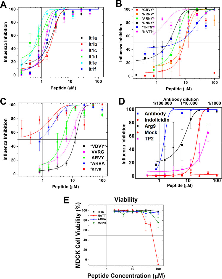
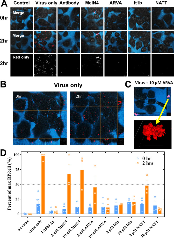
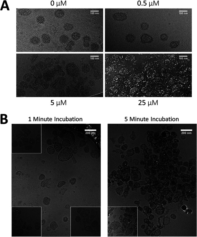
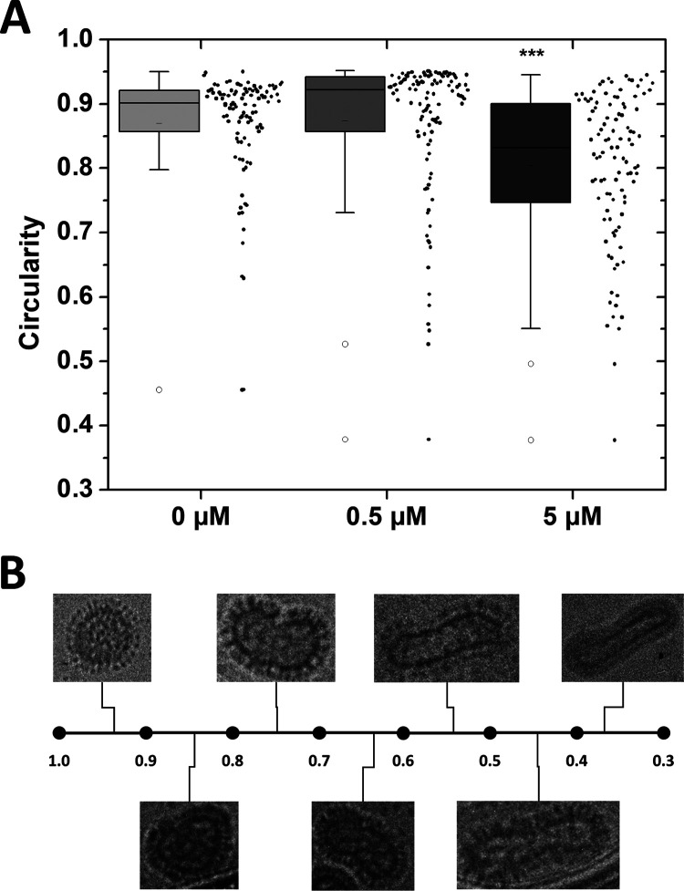
Numerous peptides inhibit the entry of enveloped viruses into cells. Some of these peptides have been shown to inhibit multiple unrelated viruses. We have suggested that such broad-spectrum antiviral peptides share a property called interfacial activity; they are somewhat hydrophobic and amphipathic, with a propensity to interact with the interfacial zones of lipid bilayer membranes. In this study, we further tested the hypothesis that such interfacial activity is a correlate of broad-spectrum antiviral activity. In this study, several families of peptides, selected for the ability to partition into and disrupt membrane integrity but with no known antiviral activity, were tested for the ability to inhibit multiple diverse enveloped viruses. These include Lassa pseudovirus, influenza virus, dengue virus type 2, herpes simplex virus 1, and nonenveloped human adenovirus 5. Various families of interfacially active peptides caused potent inhibition of all enveloped viruses tested at low and submicromolar concentrations, well below the range in which they are toxic to mammalian cells. These membrane-active peptides block uptake and fusion with the host cell by rapidly and directly interacting with virions, destabilizing the viral envelope, and driving virus aggregation and/or intervirion envelope fusion. We speculate that the molecular characteristics shared by these peptides can be exploited to enable the design, optimization, or molecular evolution of novel broad-spectrum antiviral therapeutics.IMPORTANCE New classes of antiviral drugs are needed to treat the ever-changing viral disease landscape. Current antiviral drugs treat only a small number of viral diseases, leaving many patients with established or emerging infections to be treated solely with supportive care. Recent antiviral peptide research has produced numerous membrane-interacting peptides that inhibit diverse enveloped viruses in vitro and in vivo Peptide therapeutics are becoming more common, with over 60 FDA-approved peptides for clinical use. Included in this class of therapeutics is enfuvirtide, a 36-residue peptide drug that inhibits HIV entry/fusion. Due to their broad-spectrum mechanism of action and enormous potential sequence diversity, peptides that inhibit virus entry could potentially fulfill the need for new antiviral therapeutics; however, a better understanding of their mechanism is needed for the optimization or evolution of sequence design to combat the wide landscape of viral disease.

Keywords: entry inhibitor; interfacial activity; membrane; peptide.


Figures

FIG 1
FIG 1
Peptide inhibition of LASVpv and cytotoxicity. Pseudovirus was incubated with various concentrations of peptide for 1 h and was then added to HEK 293T/17 cells. Infectivity was quantified by measuring luciferase expression translated from the LASVpv genome approximately 72 h after infection. Cells were also treated with peptide alone, and cell viability was determined by alamarBlue approximately 72 h after treatment. Infectivity is shown as solid points, and respective nonlinear curve fits are shown as solid lines. Cell viability is shown as empty points with dashed lines. Peptide libraries tested were the It1 family (A), BS family (B), and VS family (C), as well as miscellaneous and control peptides (D). Points are means ± SE; n = 4 to 8.
FIG 2
FIG 2
Inhibition of influenza virus. Inhibition of influenza A/Hong Kong/8/68 (H3N2) virus was measured for the It1 peptides (A), the BS peptides (B), the VS peptides (C), and the set of control peptides (D; see the text). We also used mock inhibition as a buffer-only positive control and used a 1:50,000 dilution of a human convalescent-phase antibody (BEI Resources) as an inhibition control. Virus at 50× TCID50 was incubated in 96-well plates with serially diluted peptide for 1 h and was then added to MDCK cells for 1 h, followed by washing and the addition of fresh media. After 48 h at 37°C, the supernatants were removed for further assays; the plates were then washed, fixed with 4% paraformaldehyde, and stained with DAPI. DAPI fluorescence enables the measurement of intact cells remaining in the well. Viral cytopathic effects destroy the cell monolayer under these conditions, unless inhibited; cells that have succumbed to infection are washed away before fixation. Points are means ± SE; n = 4 to 10. (E) Viability of MDCK cells after treatment with one representative peptides from each set of peptides measured by SYTOX green staining.
FIG 3
FIG 3
Confirmation of influenza virus growth and inhibition for some example peptides. (A) Western blotting. Aliquots of the supernatant from viral inhibition experiments (Fig. 2) were subjected to SDS-PAGE, transferred, and then blotted with an anti-hemagglutinin primary antibody and a peroxidase-labeled secondary antibody. One Western blot from the biologically selected peptides (*RNNY*) and another from the It1 family (It1f) are shown. (B) Hemagglutination assay. d-ARVA (*arva) is the d-amino acid variant of *ARVA. Example experiments were performed with infection supernatant from the inhibition assay (Fig. 2). In the top row, infection was done with virus treated with a peptide concentration of 50 μM or antibody dilution of 1/1,000. Treatments were serially diluted by a factor of 2 down the plate. Culture supernatants were mixed with 1% turkey RBCs and then incubated at room temperature for 1 h. Hemagglutination prevented the RBCs from settling into a distinct pellet. (C) Virus propagation time course. MDCK cells were infected with various titers of influenza A (H1N1) virus, untreated, or treated with peptide 30 min before or 30 min after infection. Virus propagation was measured by qPCR at multiple time points postinfection (n = 3; mean ± SE).
FIG 4
FIG 4
Peptides inhibit multiple diverse viruses. Peptides were tested against dengue virus type 2 (A), herpes simplex virus 1 (B), and human adenovirus 5 (C) (n = 6, mean ± SE). Plaque assays in Vero E6 cells were used to quantify infectivity. Virus and peptide were incubated for 1 h. The inoculum was transferred to cells for 1 h and then washed from the cells. Avicel overlay was added to cells and then incubated until viral plaques were visible by crystal violet staining. (D) Peptide cytotoxicity against Vero E6 cells was measured by SYTOX green staining.
FIG 5
FIG 5
Time-of-addition assay. Peptides, at 10 μM, were added to cells at various time points before and after LASVpv infection (0 min). Representatives of each interfacially active peptide family (A to C) were tested, as well as two controls (D). Controls include the negative peptide MelN4 and an anti-Lassa virus antibody. Infectivity was quantified approximately 72 h after infection by measuring luciferase expression translated from the LASVpv genome (n = 4; mean ± SE). Significance relative to −60 min was determined by one-way analysis of variance (ANOVA) with Dunnett’s posttest. *, P < 0.05; **, P < 0.01; ***, P < 0.001; n.s., no statistical difference. (E) Effect of preincubation with cells instead of virus. Standard series (s) was treated in the same manner as that for Fig. 1; various concentrations of It1b were first incubated with LASVpv for 1 h and then added to HEK 239T/17 cells. For the wash series (w), It1b was first incubated on cells for 1 h. Cells were then washed with PBS to remove excess unbound peptide, followed by the addition of LASVpv to the cells. Infectivity of both series was quantified by measuring luciferase expression translated from the LASVpv genome approximately 72 h after infection. Data are represented as the difference in inhibition between the two series (w − s). n = 8; mean ± SE.
FIG 6
FIG 6
Antiviral peptides prevent viral entry into mammalian cells. (A) R18-labeled H1N1 influenza virus was preincubated with peptides and then incubated with A549 cells for 1 h at 4°C to allow binding but not uptake of virus. After the incubation, cells were washed. Confocal microscopy images were taken at 0 and 2 h at 37°C. Negative and positive controls are shown in the first three columns. Peptide-treated samples are shown in the right four columns. The blue channel is dextran-Cascade Blue to show cell borders. The red is R18, which is self-quenched in intact virions and becomes fluorescent upon endosomal fusion. (B) Confocal images with depth profiles on left and lower edge. At 0 h, viral particles were bound only to the cell surface. After 2 h at 37°C, particles were largely within the cell and were more fluorescent due to virus-endosome fusion. (C) With peptide treatment, large viral aggregates were observed in the extracellular space or on the cell surface, but they did not enter cells. Scale bar = 5 μm. (D) Quantitation of viral entry and fusion. Red particles (RP) brighter than a background-level threshold that are inside treated cells were counted with ImageJ and compared to the number in untreated cells. Large particles (>1 μm) (see panel C) were excluded, based on size and also because they were always located outside the cells. The results are expressed as percentages of virus-only control. Individual experiments are shown as points. Bars are means ± SE; n = 3 to 8.
FIG 7
FIG 7
Virions rapidly form aggregates as peptide concentration increases. (A) H1N1 influenza virus was incubated with various concentrations of NATT peptide for 30 min and then inactivated by UV light. Samples were then visualized by cryo-electron microscopy. (B) UV-inactivated H1N1 was incubated with 25 μM NATT peptide for 1 and 5 min and then immediately frozen in liquid ethane. Samples were visualized by cryo-electron microscopy. Insets are shown from addition images of the same sample.
FIG 8
FIG 8
Virion morphology changes as peptide concentration increases. (A) Viral envelopes from Fig. 7A were traced, and the circularity of each virion was measured using ImageJ. Data are represented by box plot (left of x axis tick) as well as individual measurements (right of x axis tick). Significance was determined by one-way ANOVA with Tukey’s posttest. ***, P < 0.001. n = 103 to 115. (B) Representative set of virions displaying a wide range of circularity.

Similar articles

Cited by

References

    1. Heron M. 2019. National Vital Statistics Reports deaths: leading causes for 2017. Natl Vital Stat Rep 68:1–77. - PubMed
    1. Murphy SL, Xu J, Kochanek KD, Arias E. 2018. Mortality in the United States, 2017. NCHS Data Brief 2018(328):1–8. - PubMed
    1. Johnson NB, Hayes LD, Brown K, Hoo EC, Ethier KA, Centers for Disease Control and Prevention. 2014. CDC National Health Report: leading causes of morbidity and mortality and associated behavioral risk and protective factors—United States, 2005–2013. MMWR Suppl 63:3–27. - PubMed
    1. De Clercq E, Li G. 2016. Approved antiviral drugs over the past 50 years. Clin Microbiol Rev 29:695–747. doi:10.1128/CMR.00102-15. - DOI - PMC - PubMed
    1. Littler E, Oberg B. 2005. Achievements and challenges in antiviral drug discovery. Antivir Chem Chemother 16:155–168. doi:10.1177/095632020501600302. - DOI - PubMed

Publication types

LinkOut - more resources