. 2020 Oct 27;94(22):e00125-20.
doi: 10.1128/JVI.00125-20. Print 2020 Oct 27.

Subcellular Localization of MxB Determines Its Antiviral Potential against Influenza A Virus

Affiliations

Subcellular Localization of MxB Determines Its Antiviral Potential against Influenza A Virus

Fiona Steiner et al. J Virol. .

Abstract

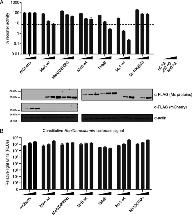
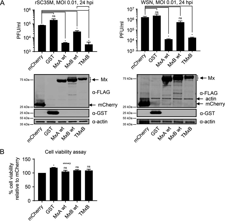
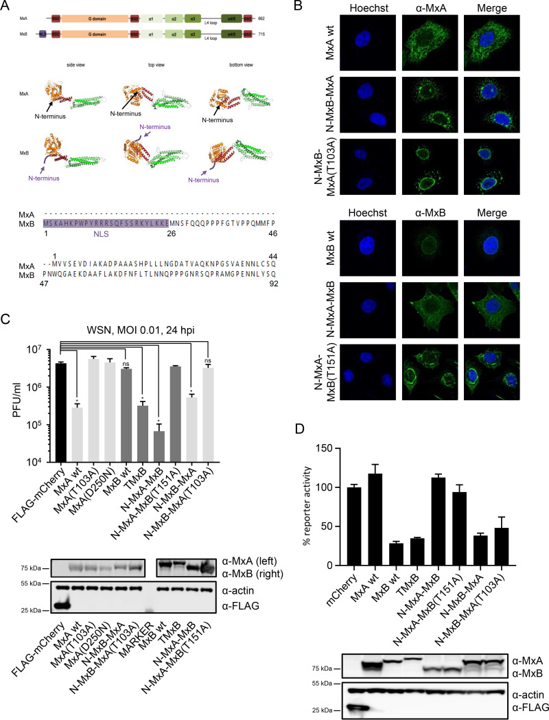
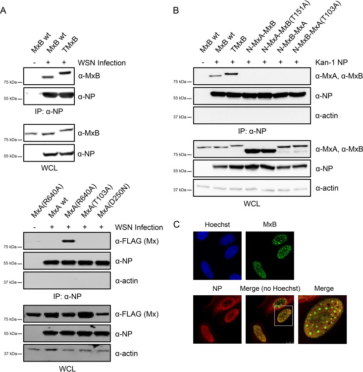
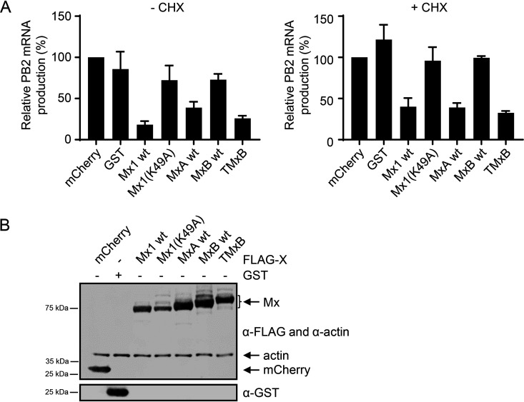
Mx proteins are interferon (IFN) type I (α/β)- and type III (λ)-induced effector proteins with intrinsic antiviral activity. Mammalian Mx proteins show different subcellular localizations and distinct yet partially overlapping viral specificities. However, the precise mechanism(s) of antiviral action are still unresolved. Human MxA accumulates in the cytoplasm and inhibits a wide variety of RNA and DNA viruses, among them influenza A virus (IAV). In contrast, MxB, the second human Mx protein, localizes via its amino (N) terminus to the outer nuclear membrane at or near nuclear pores and inhibits the nuclear import of incoming human immunodeficiency viruses (HIV) and herpesviruses, but not that of IAV. Here, we evaluated whether the antiviral specificity of MxB is determined by its subcellular localization. For this purpose, we redirected MxB to the nucleus or cytoplasm by either attaching a nuclear localization signal to its N terminus or by exchanging the N terminus of MxB with that of MxA. Interestingly, ectopic expression of these MxB variants in the nucleus or in the cytoplasm rendered the host cells resistant to IAV, revealing that the capacity of MxB to block IAV replication critically depends on the site where the protein accumulates in the infected cell. Furthermore, coimmunoprecipitation (co-IP) assays demonstrated that MxB physically interacted with the nucleoprotein (NP) of IAV. Taken together, the data indicate that the subcellular localization of the MxB protein plays a pivotal role in determining its antiviral specificity.IMPORTANCE The interferon system plays a pivotal role in the defense against viral infections. The dynamin-related Mx proteins form a small family of interferon-induced effector proteins with distinct antiviral specificities and subcellular localizations. So far, it is not clear whether the different virus specificities of Mx proteins are the result of distinct mechanisms of action or are due rather to their different subcellular localization. We show here that the human MxB protein, normally localized to the outer membrane of the cell nucleus, acquires antiviral activity against IAV when redirected to the nucleus or cytoplasm, subcellular sites where other members of the Mx protein family efficiently interfere with IAV replication. Our findings thus strongly suggest that Mx proteins act primarily through a common mechanism and that their viral specificity is at least in part determined by their individual subcellular localization.

Keywords: Mx proteins; MxB; influenza A viruses; interferon system; subcellular localization; viral specificity.


Figures

FIG 1
FIG 1
TMxB accumulates in the cell nucleus and is associated with promyelocytic leukemia (PML) bodies. (A) HEp-2 cells were transfected with plasmids coding for FLAG-tagged wild-type (wt) MxB (MxB wt), FLAG-tagged MxB containing an N-terminal nuclear localization signal (NLS), (TMxB), and FLAG-tagged wild-type mouse Mx1 (Mx1 wt). Cells were fixed and immune stained with a mouse monoclonal antibody specific for the FLAG tag. Images are representative for two independent experiments. (B) HEp-2 cells were transfected with plasmids coding for nuclear MxB (TMxB) and wild-type Mx1. Cells were fixed and immune stained with a mouse monoclonal antibody specific for the FLAG tag and with a rabbit polyclonal PML antibody. PML bodies, promyelocytic leukemia bodies in the nucleus. Arrows depict examples of TMxB-PML body in juxtaposition.
FIG 2
FIG 2
The nuclear variant of MxB (TMxB) exhibits restriction of IAV replication. (A) Minireplicon assay of 293T cells transfected with plasmids encoding the minireplicon constituents and increasing amounts of FLAG-mCherry or the indicated FLAG-Mx proteins. Cells were lysed and a dual-luciferase assay was performed. Values are presented in percent firefly luciferase activity normalized to Renilla luciferase relative to the mCherry control for each individual condition (66 ng, 200 ng, and 600 ng). Values of mCherry-expressing samples are set to 100%. Data are represented as mean ± standard deviation (SD) of triplicates. The experiment was performed in biological triplicates. Western blot analysis was carried out with pooled lysates for each condition, and immunostaining was performed with the indicated antibodies (bottom). (B) Renilla luciferase activity in the minireplicon assays shown in panel A. Data are represented as mean ± SD of triplicates.
FIG 3
FIG 3
TMxB blocks IAV infection. (A) Plaque assay titration of supernatants harvested from 293T cells transiently transfected with plasmids coding for the indicated control protein or FLAG-Mx proteins and infected with a multiplicity of infection (MOI) of 0.01 for 24 h. Supernatants were titrated on MDCK cells. Data are represented as mean ± standard error of the mean (SEM) of duplicates. Bottom panels show expression control. Samples were harvested at the same time as supernatants were collected for the plaque assay. Western blot analysis was performed using the indicated antibodies (bottom panel). Data are representative for two independent experiments. (B) CellTiter-Glo cell viability assay. 293T cells were transfected as in panel A and cells were lysed at the same time as supernatant was collected for the plaque assay from an identical plate. Luminescence was measured, and absolute values were plotted. Data are represented as mean ± SEM of duplicates. P < 0.05; Student’s t test.
FIG 4
FIG 4
MxB with the N terminus of MxA relocalizes to the cytoplasm and acquires anti-IAV activity. (A) Top: graphical representation of MxA and MxB. Middle: crystal structure of MxA (28-amino-acid deletion in loop L4; PDB identifier [ID] 3SZR) and MxB (83-amino-acid deletion at the N terminus; PDB ID 4WHJ). Bottom: alignment of the amino acid sequence of the N termini of MxA and MxB. (B) Subcellular localization of MxA-MxB chimera. HEp-2 cells were transfected with plasmids expressing the indicated MxA or MxB variants or chimera. Fixed cells were immunostained with a monoclonal mouse anti-MxA or anti-MxB antibody. Shown are representative images of 2 independent experiments. (C) IAV replication capacity in the presence of MxA, MxB, TMxB, and the indicated MxA-MxB chimera. 293T cells were transiently transfected with plasmids coding for the indicated proteins for 24 h and were subsequently infected with WSN at an MOI of 0.01 for 24 h. Protein expression was controlled by immunoblot analysis. Supernatant was transferred to MDCK cells and viral titers were determined 24 h postinfection. Data are representative for three independent experiments. Data are represented as mean ± SD of triplicates. P < 0.0001; Student’s t test. (D) HIV-1 reporter virus replication capacity in the presence of MxA, MxB, TMxB, and the indicated MxA-MxB chimera. HeLa cells were transiently transfected with expression plasmids coding for the indicated proteins. At 24 h posttransfection, cells were infected with VSV-G-pseudotyped HIV-1 NL-Luc reporter virus. Luciferase assay was performed at 48 h postinfection, and reporter activity is shown relative to mCherry. Data are represented as mean ± SD of duplicates. Data are representative for three independent experiments.
FIG 5
FIG 5
MxB forms a stable complex with NP. (A) HeLa cells were transfected with plasmids encoding the indicated Mx proteins. At 24 h posttransfection, cells were infected with WSN at an MOI of 5 for 6 h. Cells were lysed, and viral NP was immunoprecipitated (IP) from lysates using a mouse monoclonal anti-NP antibody. The resulting complexes were subjected to Western blot analysis using the indicated antibodies. In addition, whole-cell lysates (WCL) were also monitored for expression of Mx, NP, and actin by Western blot analysis. (B) HEp-2 cells were transfected with plasmids coding for the indicated Mx proteins and either a Kan1 NP-expressing plasmid or an empty vector. At 24 h posttransfection, cells were lysed and the IP was performed as in panel A. (C) HEp-2 cells were transfected with plasmids coding for FLAG-TMxB and WSN NP. Cells were fixed and stained with a rabbit antibody specific for the FLAG tag and a mouse monoclonal antibody specific for IAV NP.
FIG 6
FIG 6
Primary transcription of IAV is inhibited by TMxB. (A) 293T cells were transfected with plasmids coding for the indicated control or FLAG-Mx proteins and infected with rSC35M(H7N7) at an MOI of 5 for 6 h. RNA was isolated and reverse transcribed into cDNA using random primers. RT-qPCR analysis was performed, and PB2 mRNA normalized to GAPDH mRNA is shown. Cells were pretreated with 100 μg/ml cycloheximide (CHX) or with the same volume of solvent (DMSO) for 1 h prior to infection. Where indicated, infection was carried out in the presence of CHX. Values are relative to the mCherry control, which was set to 100%. Data are represented as mean ± SEM of triplicates. (B) Western blot analysis of 293T cells transfected and lysed at the same time as those in panel A. Immunostaining was performed with the indicated antibodies.

Similar articles

Cited by

References

    1. Haller O, Staeheli P, Schwemmle M, Kochs G. 2015. Mx GTPases: dynamin-like antiviral machines of innate immunity. Trends Microbiol 23:154–163. doi:10.1016/j.tim.2014.12.003. - DOI - PubMed
    1. Staeheli P, Haller O. 2018. Human MX2/MxB: a potent interferon-induced postentry inhibitor of herpesviruses and HIV-1. J Virol 92:e00709-18. doi:10.1128/JVI.00709-18. - DOI - PMC - PubMed
    1. Verhelst J, Hulpiau P, Saelens X. 2013. Mx proteins: antiviral gatekeepers that restrain the uninvited. Microbiol Mol Biol Rev 77:551–566. doi:10.1128/MMBR.00024-13. - DOI - PMC - PubMed
    1. Zürcher T, Pavlovic J, Staeheli P. 1992. Nuclear localization of mouse Mx1 protein is necessary for inhibition of influenza virus. J Virol 66:5059–5066. doi:10.1128/JVI.66.8.5059-5066.1992. - DOI - PMC - PubMed
    1. Zurcher T, Pavlovic J, Staeheli P. 1992. Mechanism of human MxA protein action: variants with changed antiviral properties. EMBO J 11:1657–1661. doi:10.1002/j.1460-2075.1992.tb05212.x. - DOI - PMC - PubMed

Publication types