

Cervical Myelopathy Due to Ochronosis: An Intraoperative Suspicion
- PMID: 32908119
- PMCID: PMC7491942
- DOI: 10.12659/AJCR.924575
Cervical Myelopathy Due to Ochronosis: An Intraoperative Suspicion
Abstract
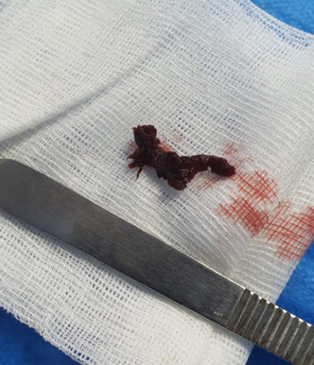
BACKGROUND Alkaptonuria (AKU) is a rare metabolic disease caused by a deficiency in homogentisic acid oxidase, which leads to the accumulation of homogentisic acid dark pigments in tissues such as bones, ligaments, and tendons. Long-term duration of this condition, termed ochronosis, can result in degenerative arthropathy involving the spine and large joints. CASE REPORT This report describes a 55-year-old Jordanian woman presenting with chronic neck and lower back pain. History, physical examination, and radiological imaging indicated cervical myelopathy and lumbar spine degeneration. Two-level anterior cervical discectomy and fusion was performed successfully. Intra-operatively, the cervical discs were observed to be black, suggesting a diagnosis of alkaptonuria, which was later confirmed by genetic testing. A detailed history and physical examination revealed the absence of classical features of AKU. CONCLUSIONS Intraoperative detection of black disc material suggests the need for further tests to diagnose AKU, especially in indolent patients who did not have classical clinical features. Surgical management may improve outcomes in patients with cervical myelopathy due to ochronosis.
Conflict of interest statement
None.
Figures


Similar articles
-
Ochronosis and lumbar disc herniation.Acta Neurochir (Wien). 2006 Aug;148(8):891-4; discussion 894. doi: 10.1007/s00701-006-0774-9. Epub 2006 May 29. Acta Neurochir (Wien). 2006. PMID: 16736091
-
[Ochronosis: A case report].Rev Med Interne. 2022 Nov;43(11):669-672. doi: 10.1016/j.revmed.2022.05.003. Epub 2022 Jun 2. Rev Med Interne. 2022. PMID: 35659777 French.
-
Not just another case of low back pain.BMJ Case Rep. 2014 Apr 29;2014:bcr2014204085. doi: 10.1136/bcr-2014-204085. BMJ Case Rep. 2014. PMID: 24781848 Free PMC article.
-
Disc-coloration of an ochronotic cervical intervertebral disc in a patient with alkaptonuria: Case report and review of the literature.Clin Neurol Neurosurg. 2024 Jul;242:108349. doi: 10.1016/j.clineuro.2024.108349. Epub 2024 May 22. Clin Neurol Neurosurg. 2024. PMID: 38820945 Review.
-
"As Black as Ink": A Case of Alkaptonuria-Associated Myelopathy and a Review of the Literature.Spine (Phila Pa 1976). 2019 Jan 1;44(1):E53-E59. doi: 10.1097/BRS.0000000000002755. Spine (Phila Pa 1976). 2019. PMID: 29933333 Review.
References
-
- Phornphutkul C, Introne WJ, Perry MB, et al. Natural history of alkaptonuria. N Engl J Med. 2002;347(26):2111–21. - PubMed
-
- Zatkova A. HGD mutation database. Oct 24, 2010. http://hgddatabase.cvtisr.sk/home.php?select_db=HGD.
-
- Donaldson CJ, Mitchell SL, Riley LH, 3rd, Kebaish KM. “As black as ink”: A case of alkaptonuria-associated myelopathy and a review of the literature. Spine (Phila Pa 1976) 2019;44(1):E53–59. - PubMed
-
- Kahveci R, Ergüngör MF, Günaydin A, Temiz A. Alkaptonuric patient presenting with “black” disc: A case report. Acta Orthop Traumatol Turc. 2013;47(2):134–38. - PubMed
Publication types
MeSH terms
LinkOut - more resources
Full Text Sources
Medical
