. 2020 Sep 9;10(1):14849.
doi: 10.1038/s41598-020-71834-w.

Bidirectional alterations in antibiotics susceptibility in Staphylococcus aureus-Pseudomonas aeruginosa dual-species biofilm

Affiliations

Bidirectional alterations in antibiotics susceptibility in Staphylococcus aureus-Pseudomonas aeruginosa dual-species biofilm

Elena Y Trizna et al. Sci Rep. .

Abstract

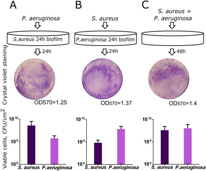
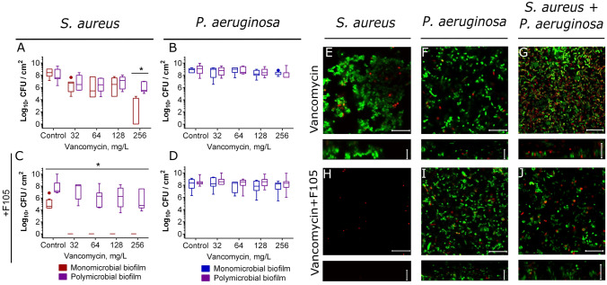
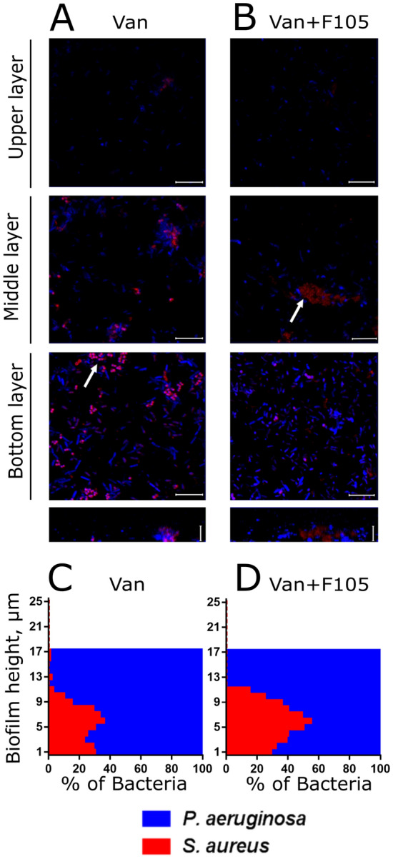
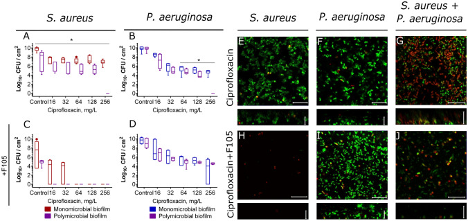
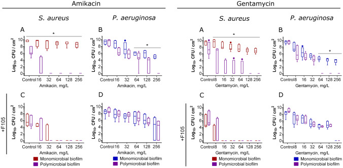
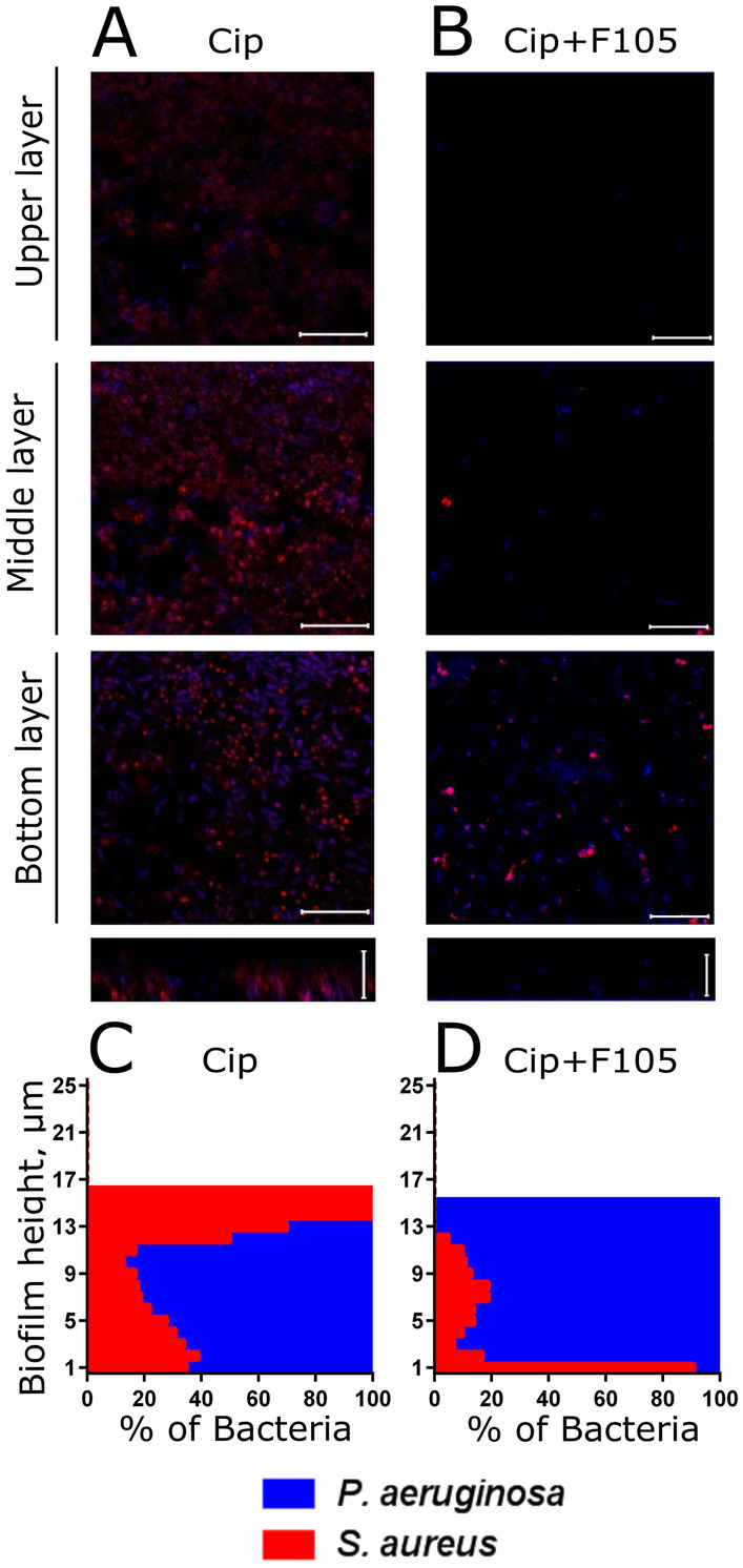
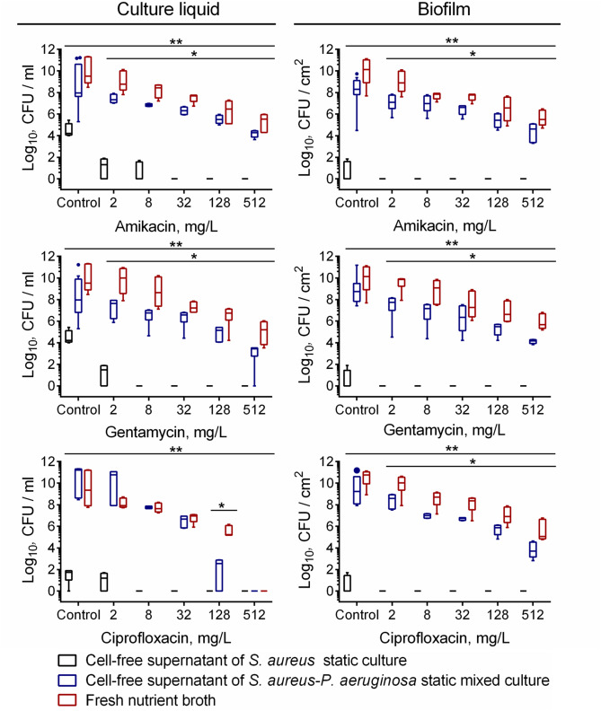
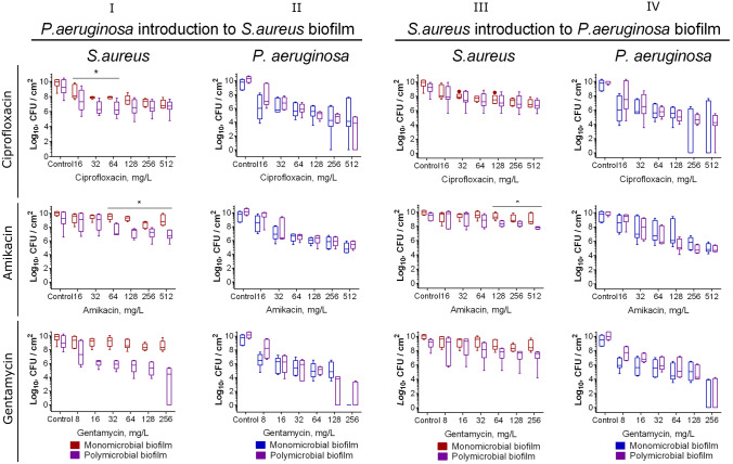
In mixed infections, the bacterial susceptibility differs significantly compared to monocultures of bacteria, and generally the concentrations of antibiotics required for the treatment increases drastically. For S. aureus and P. aeruginosa dual species biofilms, it has been numerously reported that P. aeruginosa decreases S. aureus susceptibility to a broad range of antibiotics, including beta-lactams, glycopeptides, aminoglycosides, macrolides, while sensitizes to quinolones via secretion of various metabolites. Here we show that S. aureus also modulates the susceptibility of P. aeruginosa to antibiotics in mixed cultures. Thus, S. aureus-P. aeruginosa consortium was characterized by tenfold increase in susceptibility to ciprofloxacin and aminoglycosides compared to monocultures. The same effect could be also achieved by the addition of cell-free culture of S. aureus to P. aeruginosa biofilm. Moreover, similar increase in antibiotics efficacy could be observed following addition of S. aureus suspension to the P. aeruginosa mature biofilm, compared to P. aeruginosa monoculture, and vice versa. These findings open promising perspectives to increase the antimicrobial treatment efficacy of the wounds infected with nosocomial pathogens by the transplantation of the skin residential microflora.


Conflict of interest statement

The authors declare no competing interests.

Figures

Figure 1
Figure 1
In vitro simulation of the S. aureus-P. aeruginosa mixed biofilm. (A) P.aeruginosa suspension in a fresh broth was added to the preformed 24-h old biofilm of S. aureus or (B) S. aureus was added to the preformed 24-h old biofilm of P. aeruginosa and cultivation was continued for the next 24 h. (C) As a control, both strains were inoculated simultaneously and were grown for 48 h with the broth exchange after 24 h of cultivation. The biofilms were then assessed with either crystal-violet staining of wells bottom (upper lane) or differential CFUs counting. The median values with IQRs from six independent measurements are shown.
Figure 2
Figure 2
Mono- and polymicrobial biofilms formed by S. aureus and P. aeruginosa. Cells were grown without any antimicrobial (AF) or in presence of 2(5H)-furanone derivative (F105, GL) specifically inhibiting the biofilm formation by S. aureus and exhibiting no effects on P. aeruginosa. The 48-h old biofilms were stained by Syto9/PI (AC, GI) or ViaGram Red+ (D, J) to differentiate S. aureus (stained in red) and P. aeruginosa (stained in blue) and assessed by CLSM. (E, K) The distribution of S. aureus and P. aeruginosa in the biofilm layers expressed as their relative fractions. The repression of S. aureus ica-operon in mixed biofilm by F105 was monitored by detection of GFP in ica-GFP strain (F, L). The scale bars indicate 10 µm. S. aureus microcolonies in a mixed biofilm are shown by arrows.
Figure 3
Figure 3
Atomic force microscopy (Peak Force Tapping mode) of mono- and polymicrobial biofilms formed by S. aureus and P. aeruginosa. Cells were grown without any antimicrobials (AC) or in presence of F105 specifically inhibiting the biofilm formation by S. aureus cells (DF) for 48 h, then the plates were washed, fixed with glutardialdehyde and analyzed with AFM. (I) Sensor height (topography); (II) 3D reconstruction of height channel image; (III) adhesion.
Figure 4
Figure 4
The adhesion force of S. aureus and P. aeruginosa monoculture and mixed biofilms. To repress the biofilm formation F105 was added up to 2.5 µg/ml. Asterisks denote statistical significant difference was confirmed by the Kruskal–Wallis statistical test at p < 0.05. The average values with SDs from 4 independent measurements are shown.
Figure 5
Figure 5
The effect of vancomycin on viability of S. aureus and P.aeruginosa embedded into their mono- and polymicrobial biofilms. Vancomycin was added to 48 h-old biofilms grown in absence (AB, EF) or presence (CD, HJ) of F105 to inhibit the biofilm formation by S. aureus. After 24 h incubation, the biofilms were washed twice with sterile 0.9% NaCl. The adherent cells were scratched, resuspended and CFUs were counted. The median values with IQRs from six independent measurements are shown. Asterisks denote statistical significant difference was confirmed by the Kruskal–Wallis statistical test at p < 0.05.Alternatively, biofilms were stained by Syto9/PI and biofilms were assessed by CLSM. The scale bars indicate 10 µm.
Figure 6
Figure 6
The effect of vancomycin on S. aureus and P. aeruginosa distribution in mixed biofilms. Cells were grown in absence (A, C) or in presence of F105 specifically inhibiting the biofilm formation by S.aureus cells (B, D). Vancomycin (256 µg/mL corresponding to 8 × MBC for S. aureus) was added to 48 h-old biofilms. After 24 h incubation, the biofilms were stained by ViaGram Red+ to differentiate S. aureus (stained in red) and P. aeruginosa (stained in blue) and assessed by CLSM. The scale bars indicate 10 µm. (C, D) The distribution of S. aureus and P. aeruginosa in the biofilm layers are expressed as their relative fractions.
Figure 7
Figure 7
The effect of ciprofloxacin on viability of S. aureus and P.aeruginosa embedded into their mono- and polymicrobial biofilms. Ciprofloxacin was added to 48 h-old biofilms grown in absence (AB, EF) or presence (CD, HJ) of F105 to inhibit the biofilm formation by S. aureus. After 24 h incubation, the biofilms were washed twice with sterile 0.9% NaCl. The adherent cells were scratched, resuspended and CFUs were counted. The median values with IQRs from six independent measurements are shown. Asterisks denote statistical significant difference was confirmed by the Kruskal–Wallis statistical test at p < 0.05. Alternatively, biofilms were stained by Syto9/PI and biofilms were assessed by CLSM. The scale bars indicate 10 µm.
Figure 8
Figure 8
The effect of amikacin and gentamycin on viability of S. aureus and P.aeruginosa in mono- and polymicrobial biofilms. Antimicrobials were added to 48 h-old biofilms grown in absence (AB) or presence (CD) of F105 to inhibit the biofilm formation by S. aureus. After 24 h incubation, the biofilms were washed twice with sterile 0.9% NaCl. The adherent cells were scratched, resuspended and CFUs were counted. The median values with IQRs from six independent measurements are shown. Asterisks denote statistical significant difference was confirmed by the Kruskal–Wallis statistical test at p < 0.05.
Figure 9
Figure 9
The effect of ciprofloxacin on S. aureus and P. aeruginosa distribution in mixed biofilms. Cells were grown normally (A, C) or in presence of F105 specifically inhibiting the biofilm formation by S. aureus cells (B, D). Ciprofloxacin (512 µg/mL corresponding to 8 × MBC for both bacteria) was added to 48 h-old biofilms. After 24 h incubation, the biofilms were stained by ViaGram Red+ to differentiate S. aureus (stained in red) and P. aeruginosa (stained in blue) and assessed by CLSM. The scale bars indicate 10 µm. (C, D) The distribution of S. aureus and P. aeruginosa in the biofilm layers are expressed as their relative fractions.
Figure 10
Figure 10
The effect of S. aureus and S. aureus-P.aeruginosa cell-free culture liquid on viability and susceptibility of P.aeruginosa to antimicrobials. Fresh nutrient broth (red), cells-free culture liquids of S. aureus (black) or P. aeruginosa-S. aureus mixed culture (blue) supplemented with antimicrobials in their respective 0.03–8 × MBCs were added to 48 h-old biofilms of P. aeruginosa. After 24 h incubation, the biofilms were washed twice with sterile 0.9% NaCl. The adherent cells were scratched, resuspended and CFUs were counted. The median values with IQRs from six independent measurements are shown. Asterisks and double asterisks denote statistical significant difference between fresh nutrient broth and cell-free supernatant of S. aureus culture or cell-free supernatant of S. aureus-P.aeruginosa co-culture respectively (confirmed by the Kruskal–Wallis statistical test at p < 0.05).
Figure 11
Figure 11
The susceptibility of P. aeruginosa and S. aureus after introduction of the antagonist into monoculture biofilms. (III) P. aeruginosa suspension in a fresh broth was added to the preformed 24-h old biofilm of S. aureus or (IIIIV) S. aureus was added to the preformed 24-h old biofilm of P. aeruginosa and cultivation was continued for the next 24 h. Then antimicrobials were added and after 24 h incubation CFUs in biofilms were counted. The median values with IQRs from six independent measurements are shown. Asterisks show significant difference between CFUs number between monoculture and mixed biofilms.

Similar articles

Cited by

References

    1. Kirker KR, James GA. In vitro studies evaluating the effects of biofilms on wound-healing cells: a review. Apmis. 2017;125:344–352. doi: 10.1111/apm.12678. - DOI - PubMed
    1. Kirker KR, et al. Loss of viability and induction of apoptosis in human keratinocytes exposed to Staphylococcus aureus biofilms in vitro. Wound Repair Regener. 2009;17:690–699. doi: 10.1111/j.1524-475X.2009.00523.x. - DOI - PMC - PubMed
    1. Marano RJ, et al. Secreted biofilm factors adversely affect cellular wound healing responses in vitro. Sci. Rep. 2015;5:13296. doi: 10.1038/srep13296. - DOI - PMC - PubMed
    1. Secor PR, et al. Staphylococcus aureus Biofilm and Planktonic cultures differentially impact gene expression, mapk phosphorylation, and cytokine production in human keratinocytes. BMC Microbiol. 2011 doi: 10.1186/1471-2180-11-143. - DOI - PMC - PubMed
    1. den Reijer PM, et al. Detection of alpha-toxin and other virulence factors in biofilms of Staphylococcus aureus on polystyrene and a human epidermal model. PLoS ONE. 2016 doi: 10.1371/journal.pone.0145722. - DOI - PMC - PubMed

Publication types

MeSH terms

Substances