Review
. 2021 Feb;21(2):101-115.
doi: 10.1038/s41577-020-00413-x. Epub 2020 Sep 9.

Genetic models of human and mouse dendritic cell development and function

Affiliations
Review

Genetic models of human and mouse dendritic cell development and function

David A Anderson 3rd et al. Nat Rev Immunol. 2021 Feb.

Abstract

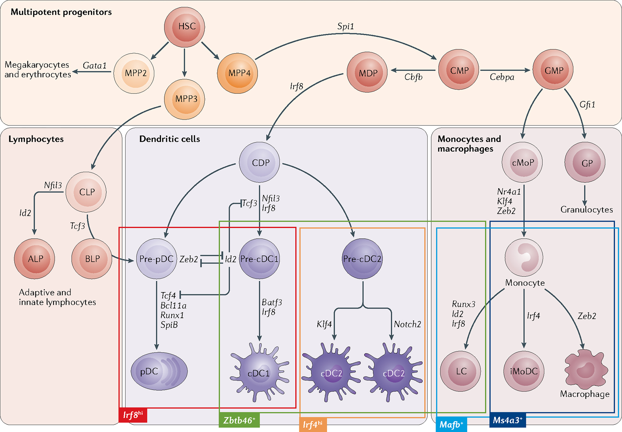
Dendritic cells (DCs) develop in the bone marrow from haematopoietic progenitors that have numerous shared characteristics between mice and humans. Human counterparts of mouse DC progenitors have been identified by their shared transcriptional signatures and developmental potential. New findings continue to revise models of DC ontogeny but it is well accepted that DCs can be divided into two main functional groups. Classical DCs include type 1 and type 2 subsets, which can detect different pathogens, produce specific cytokines and present antigens to polarize mainly naive CD8+ or CD4+ T cells, respectively. By contrast, the function of plasmacytoid DCs is largely innate and restricted to the detection of viral infections and the production of type I interferon. Here, we discuss genetic models of mouse DC development and function that have aided in correlating ontogeny with function, as well as how these findings can be translated to human DCs and their progenitors.


Conflict of interest statement

Competing interests

The authors declare no competing interests.

Figures

Fig. 1 |
Fig. 1 |. Genetic models of mouse dendritic cell development and lineage restriction.
Hierarchical models of haematopoiesis are based on the developmental deficiencies that are observed in mice with mutations in transcription factor-encoding genes; these genes are shown adjacent to the associated stages of lineage restriction and specification. The figure highlights the development of mouse dendritic cell (DC), monocyte and macrophage subsets from shared progenitors. Haematopoietic stem cell (HSC)-derived multipotent progenitors (MPPs) undergo stages of differentiation to produce lineage-restricted progenitors of lymphocytes and myeloid cells — common lymphoid progenitors (CLPs) and common myeloid progenitors (CMPs). The CLP population can be separated into two subsets on the basis of Ly6D expression. The all-lymphoid progenitor (ALP) is Ly6D and has B cell, T cell and innate lymphoid cell potential. The Tcf3-dependent B cell-biased lymphoid progenitor (BLP) is Ly6D+ and gives rise to B cells and plasmacytoid DCs (pDCs). High levels of expression of interferon regulatory factor 8 (Irf8) are associated with the specification of pDCs and type 1 classical DCs (cDC1s). Expression of Zbtb46 is a specific marker of cDC specification that is induced first in pre-cDC1s and precursor type 2 classical DCs (pre-cDC2s). cDC2 subsets are also characterized by high levels of Irf4 expression. Langerhans cells (LCs), monocytes and macrophages are all marked by Mafb-driven lineage tracing. Although LCs are not thought to be derived from the common DC precursor (CDP) population, given their embryonic origin, they do express Zbtb46 when they migrate to lymphoid organs from the skin. Lineage tracing based on Ms4a3 marks monocytes and monocyte-derived macrophages, as opposed to macrophages of embryonic origin. cMoP, common monocyte progenitor; GMP, granulocyte–macrophage progenitor; GP, granulocyte progenitor; iMoDC, immature monocyte-derived dendritic cell; MDP, monocyte–dendritic cell progenitor.
Fig. 2 |
Fig. 2 |. Stage-specific enhancer activation regulates Irf8-dependent specification of dendritic cell and monocyte progenitors.
One mechanism for the development of type 1 classical dendritic cells (cDC1s) and plasmacytoid DCs (pDCs) from bone marrow progenitors is the strict regulation of interferon regulatory factor 8 (Irf8) expression by distal, evolutionarily conserved enhancer elements. E proteins — such as TCF3 (also known as E2A) and TCF4 (also known as E2–2) — support Irf8 expression through their actions at an E-box motif-containing enhancer located 41 kb upstream of the Irf8 transcription start site. An inactivating mutation at this locus impairs Irf8 expression in pDCs and blocks the specification of precursor cDC1s (pre-cDC1s) in the common DC precursor (CDP) population. On pre-cDC1 specification, Nfil3 is induced upstream of Id2. By blocking E protein activity, ID2 imposes a requirement for an alternative enhancer located 32kb upstream of Irf8 to promote Irf8 expression. In turn, Irf8 expression and cDC1 specification are supported by BATF3 and by IRF8-dependent autoactivation. An alternative enhancer located 51kb downstream of the Irf8 locus is required for Irf8 expression regulated by PU.1 in monocytes and macrophages.
Fig. 3 |
Fig. 3 |. Specialized functions of mouse classical dendritic cell subsets.
Type 1 classical dendritic cells (cDC1s) are specialized in the regulation of type I immune responses through the priming and activation of cytotoxic CD8+ T cells and CD4+ T helper 1 (TH1) cells. Relative to other DC subsets, cDC1s specifically express Toll-like receptor 3 (TLR3) and TLR11, which recognize double-stranded RNA (dsRNA) and the Toxoplasma gondii antigen profilin, respectively. cDC1s are an essential source of IL-12 and are necessary for resistance to intracellular viral, bacterial and parasitic infections. cDC1s are uniquely capable of acquiring antigens associated with host cells, through a process known as cross-presentation, which is essential for pathogen clearance and antitumour immune responses. Type 2 classical dendritic cells (cDC2s) regulate type II and type III immune responses and antibody responses to soluble antigens. cDC2s at barrier surfaces, such as in the lung, gut and skin, regulate type II immune responses to parasites, fungi and allergens and are required for the expansion of CD4+ T helper 2 (TH2) cell populations and activation of group 2 innate lymphoid cells (ILC2s). The regulation of such responses depends on cDC-intrinsic expression of interferon regulatory factor 4 (Irf4) and has been attributed to the Klf4-dependent cDC2 subset. Type III immune responses are regulated by a distinct subset of Notch 2-dependent cDCs, which are a necessary source of IL-23 during acute infection with Citrobacter rodentium. IL-23 is necessary to activate group 3 innate lymphoid cells (ILC3s) and to induce differentiation of CD4+ T helper 17 (TH17) cells. cDC2s have also been shown to regulate antibody responses though the induction of germinal centre responses to soluble antigens in lymphoid organs. Deficiency in Notch2-dependent cDCs results in a failure to induce CD4+ T follicular helper (TFH) cells and germinal centre B cells in the spleen, for example. TCR, T cell receptor.

References

    1. Guilliams M et al. Dendritic cells, monocytes and macrophages: a unified nomenclature based on ontogeny. Nat. Rev. Immunol. 14, 571–578 (2014). - PMC - PubMed
    1. Vu Manh TP, Bertho N, Hosmalin A, Schwartz-Cornil I & Dalod M Investigating evolutionary conservation of dendritic cell subset identity and functions. Front. Immunol. 6, 260 (2015). - PMC - PubMed
    1. Reizis B Plasmacytoid dendritic cells: development, regulation, and function. Immunity 50, 37–50 (2019). - PMC - PubMed
    1. Laurenti E & Gottgens B From haematopoietic stem cells to complex differentiation landscapes. Nature 553, 418–426 (2018). - PMC - PubMed
    1. Dress RJ et al. Plasmacytoid dendritic cells develop from Ly6D+ lymphoid progenitors distinct from the myeloid lineage. Nat. Immunol. 20, 852–864 (2019). - PubMed

Publication types