. 2020 Aug 18:2020:4705768.
doi: 10.1155/2020/4705768. eCollection 2020.

Genomic and Pathogenic Characteristics of Virulent Newcastle Disease Virus Isolated from Chicken in Live Bird Markets and Backyard Flocks in Kenya

Affiliations

Genomic and Pathogenic Characteristics of Virulent Newcastle Disease Virus Isolated from Chicken in Live Bird Markets and Backyard Flocks in Kenya

Irene N Ogali et al. Int J Microbiol. .

Abstract

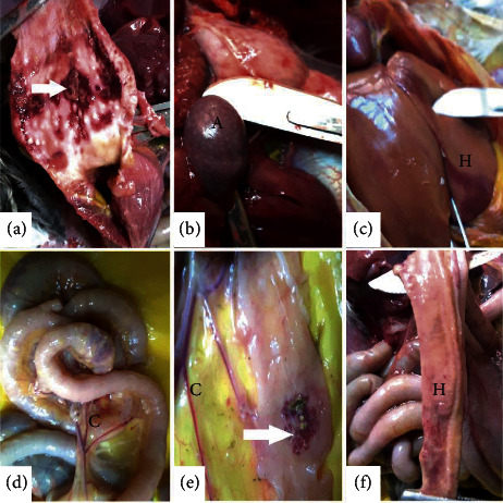
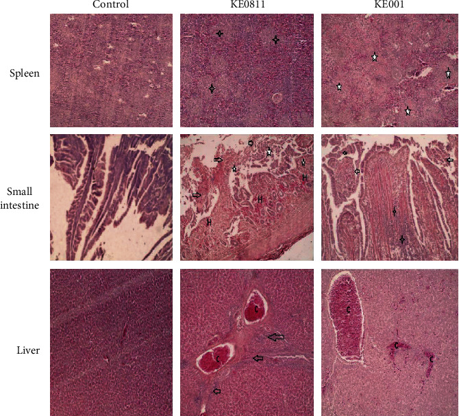
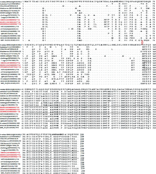
Newcastle disease (ND) causes significant economic losses in the poultry industry in developing countries. In Kenya, despite rampant annual ND outbreaks, implementation of control strategies is hampered by a lack of adequate knowledge on the circulating and outbreak causing-NDV strains. This study reports the first complete genome sequences of NDV from backyard chicken in Kenya. The results showed that all three isolates are virulent, as assessed by the mean death time (MDT) and intracerebral pathogenicity index (ICPI) in specific antibody negative (SAN) embryonated eggs and 10-day-old chickens, respectively. Also, the polybasic amino acid sequence at the fusion-protein cleavage site had the motif 112RRQKRFV118. Histopathological findings in four-week-old SPF chicken challenged with the NDV isolates KE001, KE0811, and KE0698 showed multiple organ involvement at five days after infection with severe effects seen in lymphoid tissues and blood vessels. Analysis of genome sequences obtained from the three isolates showed that they were 15192 base pair (bp) in length and had genomic features consistent with other NDV strains, the functional sites within the coding sequence being highly conserved in the sequence of the three isolates. Amino acid residues and substitutions in the structural proteins of the three isolates were similar to the newly isolated Tanzanian NDV strain (Mbeya/MT15). A similarity matrix showed a high similarity of the isolates to NDV strains of class II genotype V (89-90%) and subgenotype Vd (95-97%). Phylogenetic analysis confirmed that the three isolates are closely related to NDV genotype V strains but form a distinct cluster together with NDV strains from the East African countries of Uganda and Tanzania to form the newly characterized subgenotype Vd. Our study provides the first description of the genomic and pathological characteristics of NDV of subgenotype Vd and lays a baseline in understanding the evolutionary dynamics of NDV and, in particular, Genotype V. This information will be useful in the development of specific markers for detection of viruses of genotype V and generation of genotype matched vaccines.


Conflict of interest statement

The authors declare that there are no conflicts of interest regarding the publication of this paper.

Figures

Figure 1
Figure 1
Gross pathology lesions observed on organs of specific-pathogen-free chicken infected with NDV isolates at 5 days after challenge. First row: (a) proventriculus showed patches of reddened areas on the mucosa (hemorrhagic patches), (b) the spleen was oedematous and showed multifocal white specks (necrotic points-A), and (c) the liver was oedematous and showed focal reddened hemorrhagic areas (H). Second row: ((d–f)) small intestines showed congestion of blood vessels (C), darkened patches of necrosis (arrow), and diffuse hemorrhages (H) on the mucosa.
Figure 2
Figure 2
Photomicrographs showing hematoxylin and eosin (HE) staining on sections of the spleen, small intestines, and liver at 5 days after challenge. Magnification for panels of spleen and liver and 100x for small intestines × 400. First row: spleen tissue samples from isolate: KE0811 and KE001 showed lymphocyte depletion in the follicles and inflammatory exudates (star). Second row: small intestine tissue samples from NDV challenged groups showed multifocal mucosal hemorrhages (H), lymphocytic infiltration of the submucosa (star), and sloughing of mucosa (arrow). Third row: liver tissue samples of NDV challenged group showed congestion of hepatic blood vessels (C) and lymphocyte infiltration of periportal areas (arrow).
Figure 3
Figure 3
Alignment of the V protein of NDV isolates. The amino acid sequence of the V protein of the study isolates (highlighted in red) was aligned with NDV V proteins of each genotype reference strain. The P gene-editing site is indicated with an arrow.
Figure 4
Figure 4
Phylogenetic analysis of NDV complete genome nucleotide sequences. The evolutionary history was inferred by using the Maximum Likelihood method. The percentage of trees in which the associated taxa clustered together based on 1000 replicates is shown next to the branches. (a) The tree was constructed based on 306 nucleotide sequences of the fusion gene with a total of 1662 positions in the final dataset. Highlighted are sequences of isolates from Kenya that were collected in the study. The tree was constructed based on the GTR model with a discrete gamma distribution used to model evolutionary rate differences among sites (2 categories (+G), parameter = 0.33) [35]. (b) A phylogenetic tree constructed using 122 nucleotide sequences including most known African isolates. The tree is based on the General Time Reversible (GTR) model with a discrete gamma distribution used to model evolutionary rate differences among sites (2 categories (+G), parameter = 0.4054). Highlighted are sequences of Kenyan isolates collected in the study.

Similar articles

Cited by

References

    1. OIE. Newcastle disease: general infectious disease sheets. 2015. https://www.oie.int/fileadmin/Home/eng/Media_Center/docs/pdf/Disease_car....
    1. Ashraf A., Shah M. S. Newcastle disease: present status and future challenges for developing countries. African Journal of Microbiology Research. 2014;8(5):411–416. doi: 10.5897/ajmr2013.6540. - DOI
    1. Amarasinghe G. K., Bào Y., Basler C. F., et al. Taxonomy of the order Mononegavirales: update 2017. Archives of Virology. 2017;162(8):2493–2504. doi: 10.1007/s00705-017-3311-7. - DOI - PMC - PubMed
    1. Gogoi P., Ganar K., Kumar S. Avian paramyxovirus: a brief review. Transboundary and Emerging Diseases. 2017;64(1) doi: 10.1111/tbed.12355. - DOI - PubMed
    1. Dimitrov K. M., Ramey A. M., Qiu X., Bahl J., Afonso C. L. Temporal, geographic, and host distribution of avian paramyxovirus 1 (Newcastle disease virus) Infection, Genetics and Evolution. 2016;39:22–34. doi: 10.1016/j.meegid.2016.01.008. - DOI - PubMed

LinkOut - more resources