Review
. 2020 Aug 29:2020:8868593.
doi: 10.1155/2020/8868593. eCollection 2020.

Spatial Distributions, Characteristics, and Applications of Craniofacial Stem Cells

Affiliations
Review

Spatial Distributions, Characteristics, and Applications of Craniofacial Stem Cells

Geru Zhang et al. Stem Cells Int. .

Abstract

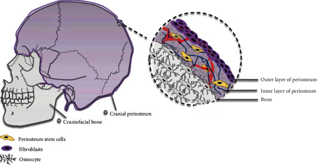
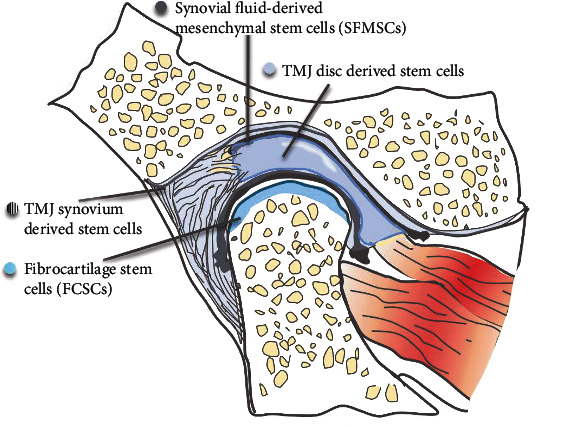
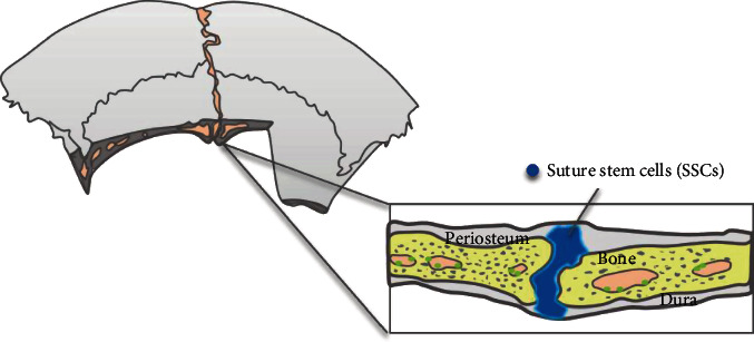
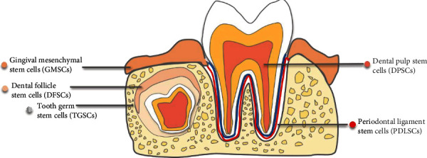
Stem cells play an irreplaceable role in the development, homeostasis, and regeneration of the craniofacial bone. Multiple populations of tissue-resident craniofacial skeletal stem cells have been identified in different stem cell niches, including the cranial periosteum, jawbone marrow, temporomandibular joint, cranial sutures, and periodontium. These cells exhibit self-renewal and multidirectional differentiation abilities. Here, we summarized the properties of craniofacial skeletal stem cells, based on their spatial distribution. Specifically, we focused on the in vivo genetic fate mapping of stem cells, by exploring specific stem cell markers and observing their lineage commitment in both the homeostatic and regenerative states. Finally, we discussed their application in regenerative medicine.


Conflict of interest statement

The authors declare that they have no competing interests.

Figures

Figure 1
Figure 1
Stem cell distribution in the cranial bone. The stem cells are located in the inner layer of calvarium periosteum.
Figure 2
Figure 2
Stem cell distribution in TMJ. Stem cells are located in the synovial fluid, synovium, disc of TMJ, and surface zone of condylar cartilage.
Figure 3
Figure 3
Stem cell distribution in sutures.
Figure 4
Figure 4
Stem cell distribution in dental tissues. Stem cells are located in gingiva, dental follicle, tooth germ, dental pulp, and periodontal ligament.

Similar articles

Cited by

References

    1. Boehm K. S., al-Taha M., Morzycki A., Samargandi O. A., al-Youha S., LeBlanc M. R. Donor site morbidities of iliac crest bone graft in craniofacial surgery: a systematic review. Annals of Plastic Surgery. 2019;83(3):352–358. doi: 10.1097/SAP.0000000000001682. - DOI - PubMed
    1. Zhang W., Yelick P. C. Craniofacial tissue engineering. Cold Spring Harbor Perspectives in Medicine. 2018;8(1) doi: 10.1101/cshperspect.a025775. - DOI - PMC - PubMed
    1. Fearon J. A., Griner D., Ditthakasem K., Herbert M. Autogenous bone reconstruction of large secondary skull defects. Plastic and Reconstructive Surgery. 2017;139(2):427–438. doi: 10.1097/PRS.0000000000002941. - DOI - PubMed
    1. Melville J. C., Mañón V. A., Blackburn C., Young S. Current methods of maxillofacial tissue engineering. Oral and Maxillofacial Surgery Clinics of North America. 2019;31(4):579–591. doi: 10.1016/j.coms.2019.07.003. - DOI - PubMed
    1. Fillingham Y., Jacobs J. Bone grafts and their substitutes. The Bone & Joint Journal. 2016;98-B(1_Supple_A):6–9. doi: 10.1302/0301-620X.98B.36350. - DOI - PubMed

LinkOut - more resources