

A Collaborative Dictionary Learning Model for Nasopharyngeal Carcinoma Segmentation on Multimodalities MR Sequences
- PMID: 32908581
- PMCID: PMC7474760
- DOI: 10.1155/2020/7562140
A Collaborative Dictionary Learning Model for Nasopharyngeal Carcinoma Segmentation on Multimodalities MR Sequences
Abstract
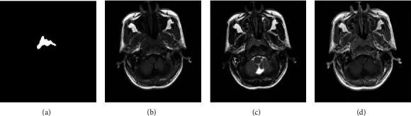
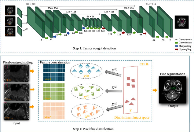
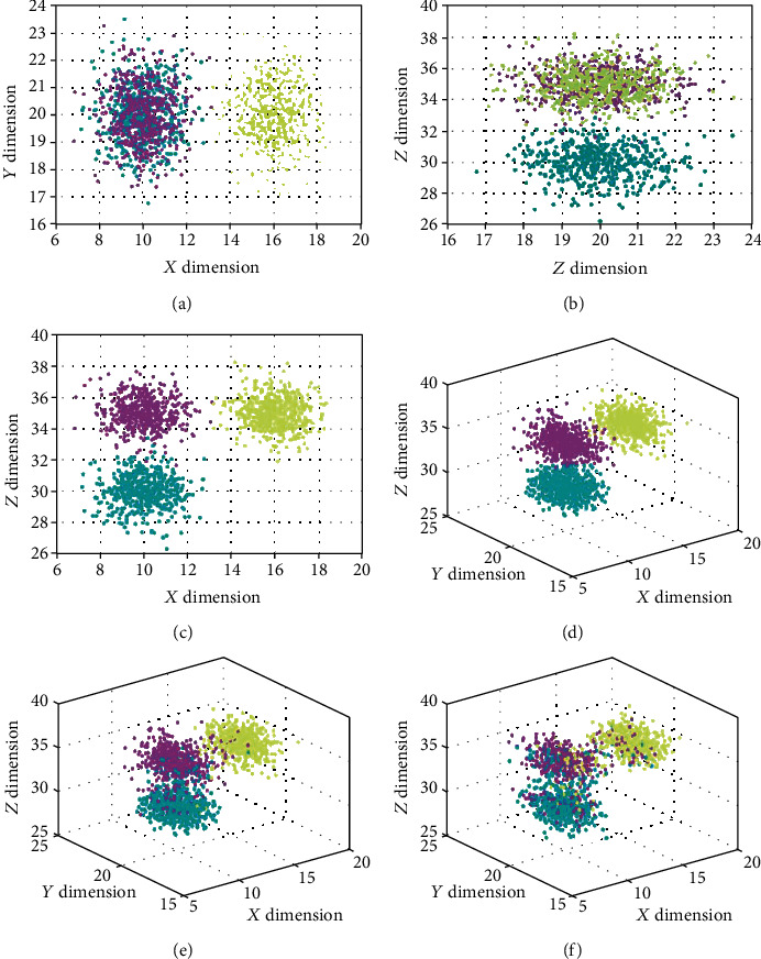
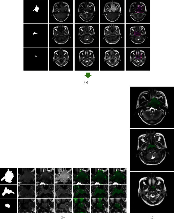
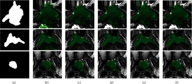
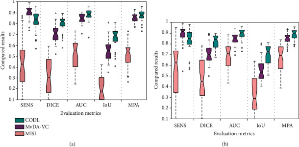
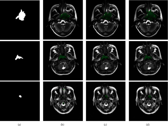
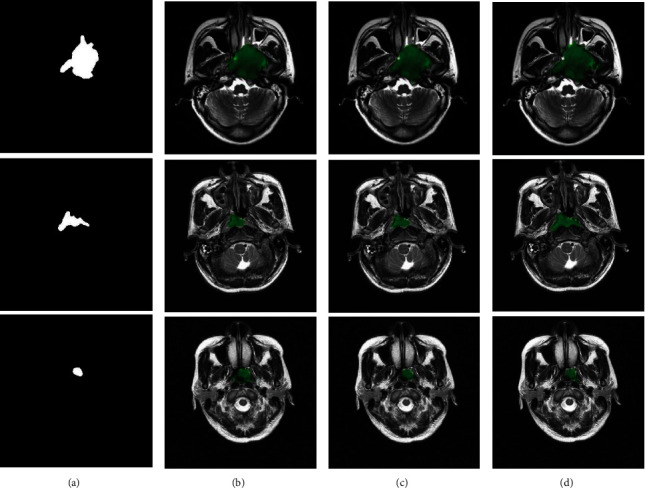
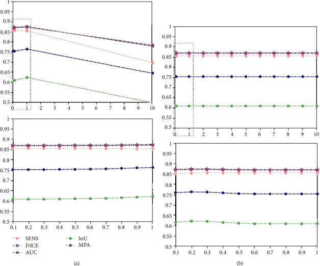
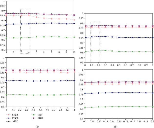
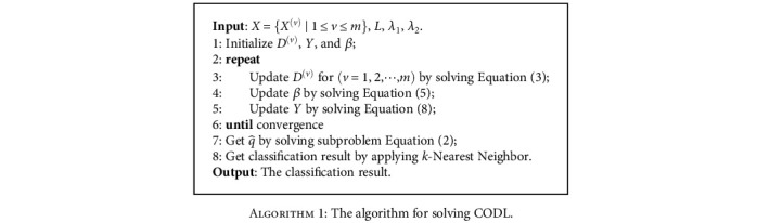
Nasopharyngeal carcinoma (NPC) is the most common malignant tumor of the nasopharynx. The delicate nature of the nasopharyngeal structures means that noninvasive magnetic resonance imaging (MRI) is the preferred diagnostic technique for NPC. However, NPC is a typically infiltrative tumor, usually with a small volume, and thus, it remains challenging to discriminate it from tightly connected surrounding tissues. To address this issue, this study proposes a voxel-wise discriminate method for locating and segmenting NPC from normal tissues in MRI sequences. The located NPC is refined to obtain its accurate segmentation results by an original multiviewed collaborative dictionary classification (CODL) model. The proposed CODL reconstructs a latent intact space and equips it with discriminative power for the collective multiview analysis task. Experiments on synthetic data demonstrate that CODL is capable of finding a discriminative space for multiview orthogonal data. We then evaluated the method on real NPC. Experimental results show that CODL could accurately discriminate and localize NPCs of different volumes. This method achieved superior performances in segmenting NPC compared with benchmark methods. Robust segmentation results show that CODL can effectively assist clinicians in locating NPC.
Copyright © 2020 Haiyan Wang et al.
Conflict of interest statement
We declare that we do not have any commercial or associative interest that represents a conflict of interest in connection with the work submitted.
Figures
Similar articles
-
SeqSeg: A sequential method to achieve nasopharyngeal carcinoma segmentation free from background dominance.Med Image Anal. 2022 May;78:102381. doi: 10.1016/j.media.2022.102381. Epub 2022 Feb 11. Med Image Anal. 2022. PMID: 35231849
-
Machine Learning Analysis of Image Data Based on Detailed MR Image Reports for Nasopharyngeal Carcinoma Prognosis.Biomed Res Int. 2020 Feb 21;2020:8068913. doi: 10.1155/2020/8068913. eCollection 2020. Biomed Res Int. 2020. PMID: 32149139 Free PMC article.
-
Radiomics on multi-modalities MR sequences can subtype patients with non-metastatic nasopharyngeal carcinoma (NPC) into distinct survival subgroups.Eur Radiol. 2019 Oct;29(10):5590-5599. doi: 10.1007/s00330-019-06075-1. Epub 2019 Mar 14. Eur Radiol. 2019. PMID: 30874880
-
A Review of Multimodal Medical Image Fusion Techniques.Comput Math Methods Med. 2020 Apr 23;2020:8279342. doi: 10.1155/2020/8279342. eCollection 2020. Comput Math Methods Med. 2020. PMID: 32377226 Free PMC article. Review.
-
Positron emission tomography/computed tomography outperforms MRI in the diagnosis of local recurrence and residue of nasopharyngeal carcinoma: An update evidence from 44 studies.Cancer Med. 2019 Jan;8(1):67-79. doi: 10.1002/cam4.1882. Epub 2018 Dec 21. Cancer Med. 2019. PMID: 30578604 Free PMC article.
Cited by
-
Prognostic potential of a voxelwise invasion risk map of nasopharyngeal carcinoma based on a coordinate system of the nasopharynx.Quant Imaging Med Surg. 2023 Feb 1;13(2):982-998. doi: 10.21037/qims-22-744. Epub 2023 Jan 5. Quant Imaging Med Surg. 2023. PMID: 36819252 Free PMC article.
-
Evolutionary route of nasopharyngeal carcinoma metastasis and its clinical significance.Nat Commun. 2023 Feb 4;14(1):610. doi: 10.1038/s41467-023-35995-2. Nat Commun. 2023. PMID: 36739462 Free PMC article.
References
MeSH terms
LinkOut - more resources
Full Text Sources
Medical
