. 2020 Aug 24;9(9):37.
doi: 10.1167/tvst.9.9.37. eCollection 2020 Aug.

Functional Analysis of Rare Genetic Variants in Complement Factor I (CFI) using a Serum-Based Assay in Advanced Age-related Macular Degeneration

Affiliations

Functional Analysis of Rare Genetic Variants in Complement Factor I (CFI) using a Serum-Based Assay in Advanced Age-related Macular Degeneration

Anuja Java et al. Transl Vis Sci Technol. .

Abstract

Purpose: Factor I (FI) is a serine protease regulator of the complement system. Genetic variants in CFI are associated with advanced age-related macular degeneration (AAMD). However, the clinical and functional impact of these variants is unknown. This study assessed the functional significance of rare CFI variants using a serum-based assay.

Methods: Carriers of rare variants with (n = 78) and without AAMD (n = 28), and noncarriers with (n = 49) and without AMD (n = 44) were evaluated. Function of FI was determined by measuring the proteolytic cleavage of C3b to iC3b, using the cofactor protein, Factor H.

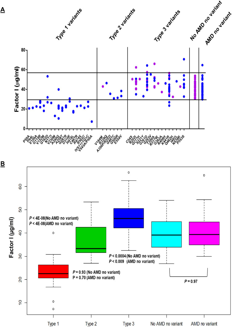
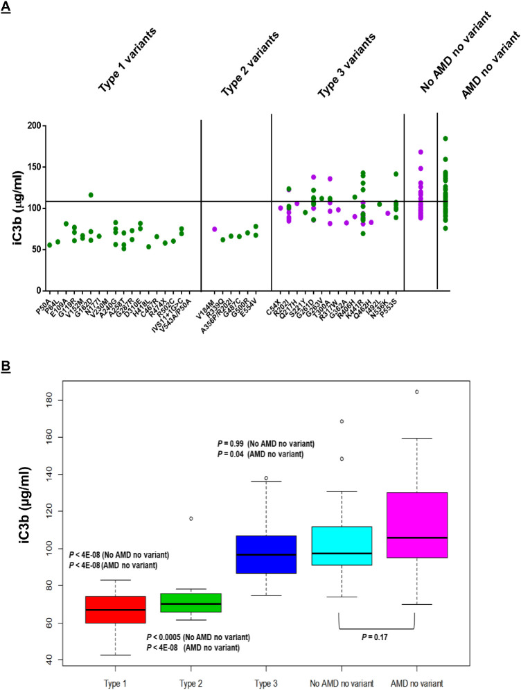
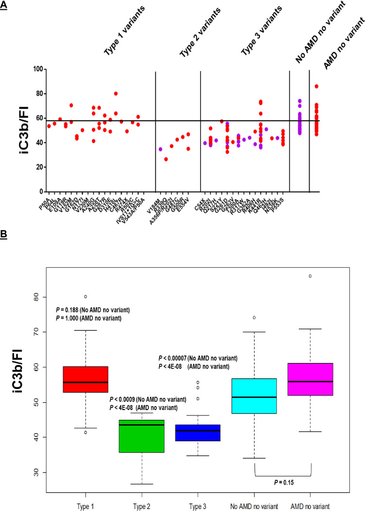
Results: CFI variants were categorized into three groups based on antigenic and functional assessments. Type 1 variants (n = 18) in 35 patients with AAMD demonstrated low serum FI levels and a corresponding decrease in FI function. Type 2 variants (n = 6) in 7 individuals demonstrated normal serum FI antigenic levels but reduced degradation of C3b to iC3b. Type 3 variants (n = 15) in 64 individuals demonstrated normal antigenic levels and degradation of C3b to iC3b. However, iC3b generation was low when measured per unit of FI. Thus most rare CFI variants demonstrate either low antigenic levels (type 1) or normal levels but reduced function (types 2 or 3).

Conclusions: Results provide for the first time a comprehensive functional assessment in serum of CFI rare genetic variants and further establish FI's key role in the pathogenesis of AAMD.

Translational relevance: Stratifying patients in the clinic with a rare CFI variant will facilitate screening and targeting patients most likely to benefit from complement therapies.

Keywords: Factor I; age-related macular degeneration; complement; functional analysis; rare genetic variants.


Conflict of interest statement

Disclosures: A. Java, Alexion Pharmaceuticals (C), Gemini Therapeutics (C), Novartis Pharmaceuticals (C); P. Baciu, Allergan (E); R. Widjajahakim, None; Y.J. Sung, None; J. Yang, Allergan (E); D. Kavanagh, Gyroscope Therapeutics (S), Alexion Pharmaceuticals (C), Sarepta (C), Actelion (C), Novartis (C), Apellis (C); J. Atkinson, Compliment Corporation (I), Kypha (I,C), Gemini Therapeutics (I), Q32BIO INC - formerly AdMiRx (I,C), Celldex Therapeutics (C), Clinical Pharmacy Services (C), Achillion Pharmaceuticals (C), BioMarin Pharmaceutical (C), Annexon Biosciences (C); J. Seddon, Laboratoires THEA (C), Gemini Therapeutics, Inc (F)

Figures

Figure 1.
Figure 1.
FI antigenic levels. (A) Individual values are shown as solid blue circles for patients with AAMD and purple circles for individuals without AAMD. Normal range (29.3–58.5 µg/mL) is represented by the black horizontal lines. The rare genetic variants are listed on the X-axis. The numbering system includes the signal peptide of 18 amino acids. (B) Box plots demonstrate that Type 1 variants have low FI antigenic levels (P < 4E-08, compared with no AMD and no variant; P < 4E-08, compared to AMD and no variant) whereas type 2 (P = 0.93, compared with no AMD and no variant; P = 0.70, compared with AMD and no variant) and type 3 variants have normal to high antigenic levels compared with controls (P < 0.0004, compared with no AMD and no variant; P < 0.009 compared to AMD and no variant). Controls (noncarriers of rare variants, with and without AMD), demonstrate normal antigenic levels of FI.
Figure 2.
Figure 2.
The iC3b generation. FI functional activity is ascertained by the quantity of iC3b generated as determined by ELISA. (A) Individual values are shown as solid green circles for AAMD and purple circles for those without AAMD. Black horizontal line represents mean iC3b value for controls. (B) Box plots demonstrate that both type 1 (P < 4E-08, compared with no AMD and no variant; P < 4E-08, compared with AMD and no variant) and type 2 variants (P < 0.0005, compared with no AMD and no variant; P < 4E-08, compared with AMD and no variant) have decreased iC3b generation compared to controls. Type 3 variants portray normal iC3b generation (P = 0.99, compared with no AMD and no variant; with P = 0.04, compared with AMD and no variant). Noncarriers (with and without AMD) also demonstrate normal iC3b generation.
Figure 3.
Figure 3.
The iC3b generation per antigenic unit (20 µg/mL) of FI. (A) Individual values are represented by solid red circles for patients with AAMD and purple circles for those without AAMD. Black horizontal line represents mean iC3b/FI for controls (noncarriers of rare variants with and without AMD). (B) Box plots demonstrate that Type 1 variants have a normal iC3b per unit of FI (P = 0.188, compared to no AMD and no variant; P = 1.00, compared with AMD and no variant). This is consistent with variants that result in expression by one allele (haploinsufficiency) of a functionally normal protein. Type 2 (P < 0.0009, compared to no AMD and no variant; P < 4E-08, compared to AMD and no variant) and type 3 variants (P < 0.00007, compared to no AMD and no variant; P < 4E-08, compared to AMD and no variant) demonstrate decreased iC3b per unit of FI since these variants result in a protein that is secreted normally but has decreased function. Noncarriers have normal iC3b generation/FI.

References

    1. Wong WL, Su X, Li X, et al.. Global prevalence of age-related macular degeneration and disease burden projection for 2020 and 2040: a systematic review and meta-analysis. Lancet Glob Health. 2014; 2: e106–e116. - PubMed
    1. Seddon JM. Macular degeneration epidemiology: nature-nurture, lifestyle factors, genetic risk, and gene-environment interactions - The Weisenfeld Award Lecture. Invest Ophthalmol Vis Sci. 2017; 58: 6513–28. - PMC - PubMed
    1. Schramm EC, Clark SJ, Triebwasser MP, Raychaudhuri S, Seddon JM, Atkinson JP. Genetic variants in the complement system predisposing to age-related macular degeneration: a review. Mol Immunol. 2014; 61: 118–25. - PMC - PubMed
    1. Geerlings MJ, de Jong EK, den Hollander AI. The complement system in age-related macular degeneration: A review of rare genetic variants and implications for personalized treatment. Mol Immunol. 2017; 84: 65–76. - PMC - PubMed
    1. Liszewski MK, Java A, Schramm EC, Atkinson JP. Complement dysregulation and disease: insights from contemporary genetics. Annu Rev Pathol. 2017; 12: 25–52. - PMC - PubMed

Publication types