

An Antibacterial Strategy of Mg-Cu Bone Grafting in Infection-Mediated Periodontics
- PMID: 32908908
- PMCID: PMC7474743
- DOI: 10.1155/2020/7289208
An Antibacterial Strategy of Mg-Cu Bone Grafting in Infection-Mediated Periodontics
Abstract
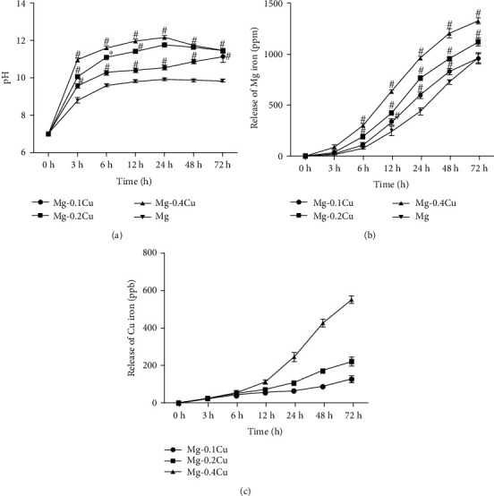
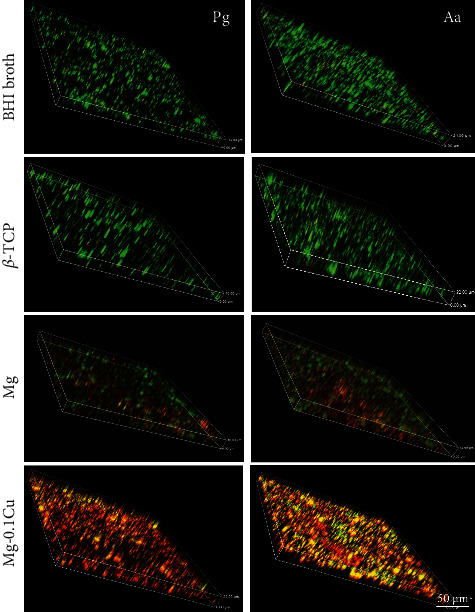
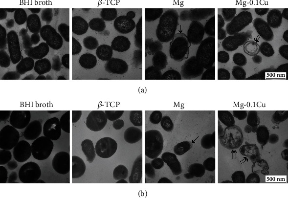
Periodontal diseases are mainly the results of infections and inflammation of the gum and bone that surround and support the teeth. In this study, the alveolar bone destruction in periodontitis is hypothesized to be treated with novel Mg-Cu alloy grafts due to their antimicrobial and osteopromotive properties. In order to study this new strategy using Mg-Cu alloy grafts as a periodontal bone substitute, the in vitro degradation and antibacterial performance were examined. The pH variation and Mg2+ and Cu2+ release of Mg-Cu alloy extracts were measured. Porphyromonas gingivalis (P. gingivalis) and Aggregatibacter actinomycetemcomitans (A. actinomycetemcomitans), two common bacteria associated with periodontal disease, were cultured in Mg-Cu alloy extracts, and bacterial survival rate was evaluated. The changes of bacterial biofilm and its structure were revealed by scanning electron microscopy (SEM) and transmission electronic microscopy (TEM), respectively. The results showed that the Mg-Cu alloy could significantly decrease the survival rates of both P. gingivalis and A. actinomycetemcomitans. Furthermore, the bacterial biofilms were completely destroyed in Mg-Cu alloy extracts, and the bacterial cell membranes were damaged, finally leading to bacterial apoptosis. These results indicate that the Mg-Cu alloy can effectively eliminate periodontal pathogens, and the use of Mg-Cu in periodontal bone grafts has a great potential to prevent infections after periodontal surgery.
Copyright © 2020 Xue Zhao et al.
Conflict of interest statement
The authors declare that they have no conflicts of interest.
Figures


Similar articles
-
Antimicrobial activity of red wine and oenological extracts against periodontal pathogens in a validated oral biofilm model.BMC Complement Altern Med. 2019 Jun 21;19(1):145. doi: 10.1186/s12906-019-2533-5. BMC Complement Altern Med. 2019. PMID: 31226983 Free PMC article.
-
Selected dietary (poly)phenols inhibit periodontal pathogen growth and biofilm formation.Food Funct. 2015 Mar;6(3):719-29. doi: 10.1039/c4fo01087f. Food Funct. 2015. PMID: 25585200
-
Antibacterial effect of copper-bearing titanium alloy (Ti-Cu) against Streptococcus mutans and Porphyromonas gingivalis.Sci Rep. 2016 Jul 26;6:29985. doi: 10.1038/srep29985. Sci Rep. 2016. PMID: 27457788 Free PMC article.
-
Oral ecology and person-to-person transmission of Actinobacillus actinomycetemcomitans and Porphyromonas gingivalis.Periodontol 2000. 1999 Jun;20:65-81. doi: 10.1111/j.1600-0757.1999.tb00158.x. Periodontol 2000. 1999. PMID: 10522223 Review.
-
Systemic antibiotics in the treatment of periodontal disease.Periodontol 2000. 2002;28:106-76. doi: 10.1034/j.1600-0757.2002.280106.x. Periodontol 2000. 2002. PMID: 12013339 Review. No abstract available.
Cited by
-
Synthesis and Characterization of Novel Sprayed Ag-Doped Quaternary Cu2MgSnS4 Thin Film for Antibacterial Application.Nanomaterials (Basel). 2022 Oct 3;12(19):3459. doi: 10.3390/nano12193459. Nanomaterials (Basel). 2022. PMID: 36234587 Free PMC article.
-
Characterization and morphological methods for oral biofilm visualization: where are we nowadays?AIMS Microbiol. 2024 Jun 6;10(2):391-414. doi: 10.3934/microbiol.2024020. eCollection 2024. AIMS Microbiol. 2024. PMID: 38919718 Free PMC article. Review.
-
Zinc alloy-based bone internal fixation screw with antibacterial and anti-osteolytic properties.Bioact Mater. 2021 May 18;6(12):4607-4624. doi: 10.1016/j.bioactmat.2021.05.023. eCollection 2021 Dec. Bioact Mater. 2021. PMID: 34095620 Free PMC article.
-
Mg-, Zn-, and Fe-Based Alloys With Antibacterial Properties as Orthopedic Implant Materials.Front Bioeng Biotechnol. 2022 May 23;10:888084. doi: 10.3389/fbioe.2022.888084. eCollection 2022. Front Bioeng Biotechnol. 2022. PMID: 35677296 Free PMC article. Review.
-
From Simple Mouth Cavities to Complex Oral Mucosal Disorders-Curcuminoids as a Promising Therapeutic Approach.ACS Pharmacol Transl Sci. 2021 Mar 17;4(2):647-665. doi: 10.1021/acsptsci.1c00017. eCollection 2021 Apr 9. ACS Pharmacol Transl Sci. 2021. PMID: 33860191 Free PMC article. Review.
References
-
- Zhu B., Macleod L. C., Newsome E., Liu J., Xu P. Aggregatibacter actinomycetemcomitans mediates protection of Porphyromonas gingivalis from Streptococcus sanguinis hydrogen peroxide production in multi-species biofilms. Scientific Reports. 2019;9(1):p. 4944. doi: 10.1038/s41598-019-41467-9. - DOI - PMC - PubMed
MeSH terms
Substances
Associated data
LinkOut - more resources
Full Text Sources
Medical
Molecular Biology Databases
