

Osteopontin Regulates Endometrial Stromal Cell Migration in Endometriosis through the PI3K Pathway : Osteopontin Regulates Endometrial Cell Migration in Endometriosis
- PMID: 32909189
- PMCID: PMC7808973
- DOI: 10.1007/s43032-020-00301-8
Osteopontin Regulates Endometrial Stromal Cell Migration in Endometriosis through the PI3K Pathway : Osteopontin Regulates Endometrial Cell Migration in Endometriosis
Abstract
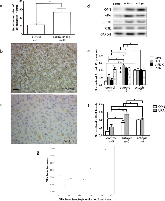
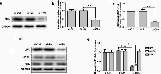
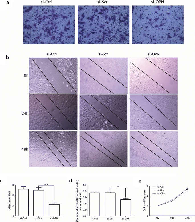
Endometriosis is generally characterized as a tumor-like disease because of its potential for distant metastasis and local tissue invasion, while whether osteopontin (OPN) plays a role in the pathogenesis of endometriosis has not been thoroughly investigated. We investigated the expression of OPN, urokinase plasminogen activator (uPA), phosphatidylinositol 3 kinase (PI3K), and phospho-PI3 kinase (p-PI3K) in endometrial stromal cells (ESCs). The serum concentration of OPN was determined by enzyme-linked immunosorbent assays (ELISA). OPN was downregulated to explore the corresponding change of uPA, p-PI3K, F-actin, and α-tubulin. The expression of OPN, uPA, PI3K, and p-PI3K was evaluated by western blot and quantitative real-time PCR (RT-qPCR) and the expression of F-actin and α-tubulin was confirmed by immunofluorescence assay. The proliferation and migration abilities of ESCs were investigated by CCK8, transwell, and wound scratch assays. Endometrial OPN, p-PI3K, and uPA expressions and serum OPN levels were increased in patients with endometriosis compared with the control. The expressions of p-PI3K, uPA, and α-tubulin were decreased by siRNA-OPN interference in ectopic ESCs. Activation and inhibition of the PI3K pathway apparently upregulate and downregulate uPA expression. Knockdown of OPN and inhibition of the PI3K pathway remarkably inhibited cell migration in ectopic ESCs. Meanwhile, activation of the PI3K pathway promoted the migration ability of ectopic ESCs. OPN may regulate the expression of uPA through the PI3K signal pathway to affect the migration ability of ESCs, indicating that OPN, uPA, and the PI3K pathway may be potential targets for interrupting development of endometriosis.
Keywords: Osteopontin; PI3K; cell migration; endometriosis; uPA.
Conflict of interest statement
The authors declare that they have no conflicts of interest.
Figures


Similar articles
-
Periostin Enhances Migration, Invasion, and Adhesion of Human Endometrial Stromal Cells Through Integrin-Linked Kinase 1/Akt Signaling Pathway.Reprod Sci. 2015 Sep;22(9):1098-106. doi: 10.1177/1933719115572481. Epub 2015 Mar 9. Reprod Sci. 2015. PMID: 25759370
-
The involvement of osteopontin and matrix metalloproteinase- 9 in the migration of endometrial epithelial cells in patients with endometriosis.Reprod Biol Endocrinol. 2015 Aug 20;13:95. doi: 10.1186/s12958-015-0090-4. Reprod Biol Endocrinol. 2015. PMID: 26289107 Free PMC article.
-
CD26/DPPIV down-regulation in endometrial stromal cell migration in endometriosis.Fertil Steril. 2014 Jul;102(1):167-177.e9. doi: 10.1016/j.fertnstert.2014.04.001. Epub 2014 May 10. Fertil Steril. 2014. PMID: 24825423
-
Osteopontin: it's role in regulation of cell motility and nuclear factor kappa B-mediated urokinase type plasminogen activator expression.IUBMB Life. 2005 Jun;57(6):441-7. doi: 10.1080/15216540500159424. IUBMB Life. 2005. PMID: 16012053 Review.
-
Osteopontin in cancer.Adv Clin Chem. 2024;118:87-110. doi: 10.1016/bs.acc.2023.11.002. Epub 2024 Jan 6. Adv Clin Chem. 2024. PMID: 38280808 Review.
Cited by
-
Osteopontin Splicing Isoforms Contribute to Endometriotic Proliferation, Migration, and Epithelial-Mesenchymal Transition in Endometrial Epithelial Cells.Int J Mol Sci. 2022 Dec 5;23(23):15328. doi: 10.3390/ijms232315328. Int J Mol Sci. 2022. PMID: 36499654 Free PMC article.
-
Effects of Progestin on Modulation of the Expression of Biomarkers in Endometriosis.Biomedicines. 2023 Jul 20;11(7):2036. doi: 10.3390/biomedicines11072036. Biomedicines. 2023. PMID: 37509675 Free PMC article.
-
TIM-3 regulates the proliferation by BDNF-mediated PI3K/AKT axis in the process of endometriosis.Mol Med. 2023 Dec 19;29(1):170. doi: 10.1186/s10020-023-00768-6. Mol Med. 2023. PMID: 38114892 Free PMC article.
-
LncRNA BANCR Promotes Endometrial Stromal Cell Proliferation and Invasion in Endometriosis via the miR-15a-5p/TRIM59 Axis.Int J Genomics. 2022 Oct 10;2022:9083822. doi: 10.1155/2022/9083822. eCollection 2022. Int J Genomics. 2022. PMID: 36262826 Free PMC article.
-
SMURF1-mediated ubiquitylation of SHP-1 promotes cell proliferation and invasion of endometrial stromal cells in endometriosis.Ann Transl Med. 2021 Mar;9(5):362. doi: 10.21037/atm-20-2897. Ann Transl Med. 2021. PMID: 33842583 Free PMC article.
References
-
- Halme J, Hammond MG, Hulka JF, Raj SG, Talbert LM. Retrograde menstruation in healthy women and in patients with endometriosis. Obstet Gynecol. 1984;64(2):151–154. - PubMed
-
- Cramer DW, Missmer SA. The epidemiology of endometriosis. Ann N Y Acad Sci. 2002;955:11–22. - PubMed
-
- Hapangama DK, Turner MA, Drury JA, Martin-Ruiz C, Von Zglinicki T, Farquharson RG, et al. Endometrial telomerase shows specific expression patterns in different types of reproductive failure. Reprod BioMed Online. 2008;17(3):416–424. - PubMed
Publication types
MeSH terms
Substances
LinkOut - more resources
Full Text Sources
Medical
Research Materials
Miscellaneous
