. 2021 Aug;35(8):4399-4416.
doi: 10.1007/s00464-020-07940-7. Epub 2020 Sep 9.

Motion analysis for better understanding of psychomotor skills in laparoscopy: objective assessment-based simulation training using animal organs

Affiliations

Motion analysis for better understanding of psychomotor skills in laparoscopy: objective assessment-based simulation training using animal organs

Koki Ebina et al. Surg Endosc. 2021 Aug.

Erratum in

Abstract

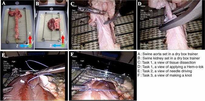
Background: Our aim was to characterize the motions of multiple laparoscopic surgical instruments among participants with different levels of surgical experience in a series of wet-lab training drills, in which participants need to perform a range of surgical procedures including grasping tissue, tissue traction and dissection, applying a Hem-o-lok clip, and suturing/knotting, and digitize the level of surgical competency.

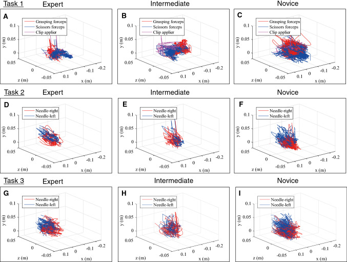
Methods: Participants performed tissue dissection around the aorta, dividing encountered vessels after applying a Hem-o-lok (Task 1), and renal parenchymal closure (Task 2: suturing, Task 3: suturing and knot-tying), using swine cadaveric organs placed in a box trainer under a motion capture (Mocap) system. Motion-related metrics were compared according to participants' level of surgical experience (experts: 50 ≤ laparoscopic surgeries, intermediates: 10-49, novices: 0-9), using the Kruskal-Wallis test, and significant metrics were subjected to principal component analysis (PCA).

Results: A total of 15 experts, 12 intermediates, and 18 novices participated in the training. In Task 1, a shorter path length and faster velocity/acceleration/jerk were observed using both scissors and a Hem-o-lok applier in the experts, and Hem-o-lok-related metrics markedly contributed to the 1st principal component on PCA analysis, followed by scissors-related metrics. Higher-level skills including a shorter path length and faster velocity were observed in both hands of the experts also in tasks 2 and 3. Sub-analysis showed that, in experts with 100 ≤ cases, scissors moved more frequently in the "close zone (0 ≤ to < 2.0 cm from aorta)" than those with 50-99 cases.

Conclusion: Our novel Mocap system recognized significant differences in several metrics in multiple instruments according to the level of surgical experience. "Applying a Hem-o-lok clip on a pedicle" strongly reflected the level of surgical experience, and zone-metrics may be a promising tool to assess surgical expertise. Our next challenge is to give completely objective feedback to trainees on-site in the wet-lab.

Keywords: Laparoscopic surgery; Motion capture; Simulation training; Surgical education.


Conflict of interest statement

Koki Ebina, Takashige Abe, Madoka Higuchi, Jun Furumido, Naoya Iwahara, Masafumi Kon, Kiyohiko Hotta, Shunsuke Komizunai, Yo Kurashima, Hiroshi Kikuchi, Ryuji Matsumoto, Takahiro Osawa, Sachiyo Murai, Teppei Tsujita, Kazuya Sase, Xiaoshuai Chen, Atsushi Konno and Nobuo Shinohara have no conflicts of interest or financial ties to disclose.

Figures

Fig. 1
Fig. 1
Photographs of the swine organ training model. A Swine aorta set in a dry box trainer. B Swine kidney set in a dry box trainer. C Task 1, a view of tissue dissection. D Task 1, a view of applying a Hem-o-lok. E Task 2, a view of needle driving. F Task 3, a view of making a knot
Fig. 2
Fig. 2
Photographs of the simulation training. A The Mocap system, which consisted of six infrared cameras (OptiTrack Prime 41, NaturalPoint Inc., USA), simultaneously tracked the movements of multiple surgical instruments during a series of training steps. B grasping forceps, C scissors. Infrared reflective marker sets with an individual arrangement pattern were attached to handles of grasping forceps, scissors, a clip applier, and needle holders. D computer display showing the movements of surgical devices. The tip movements were calculated based on the positional relationship between the tip and handle
Fig. 3
Fig. 3
Box plots of path length, velocity, acceleration, and jerk of scissors and the Hem-o-lok clip applier in Task 1 (E experts, I intermediates, N = novices). There were significant differences (p < 0.05) in the path length, velocity, acceleration, and jerk among the three groups using both scissors and the Hem-o-lok clip applier, showing the superiority of speed-related parameters and economic movements (shorter path length) of surgical devices managed by the right hand in the more experienced group. Outlier box plots represent the median (center line) and 25th and 75th percentiles (box), and the ends of the whiskers are the outermost data points from their respective quartiles that fall within the distance computed as 1.5 times the interquartile range (IQR). E experts, I intermediates, N novices
Fig. 4
Fig. 4
Representative results of trajectory of the instrument tip of an expert, an intermediate, and a novice in the three tasks. The coordinate axis is defined in Fig. 1A and B, and the origin of the coordinate is the starting point of a target object recorded at the beginning of training (see definition of "Distribution of working area" in "Materials and methods"). Overall, a shorter path length was observed in the expert group
Fig. 5
Fig. 5
Bar charts showing the distribution of the median velocity for each instrument among the three groups (E experts, I intermediates, N = novices). Overall, there was a trend toward a shorter idle state and longer state of the higher velocity range in the expert group for all instruments except the Croce grasping forceps used in Task 1. Proportion less than 1% of the very high-velocity range was not described in the figure. *, †, and ‡ indicates statistically significant (p < 0.05) among the 3 groups.  E experts, I intermediates, N = novices
Fig. 6
Fig. 6
Sub-analysis of the velocity range of the Hem-o-lok clip applier according to the working area (E experts, I intermediates, N = novices). A shorter idle state and quicker movements were significantly observed in the “near zone. Proportion less than 1% of the very high-velocity range was not described in the figure. *, †, and ‡ indicates statistically significant (p < 0.05) among the 3 groups.  E experts, I intermediates, N = novices
Fig. 7
Fig. 7
Principal component analysis regarding the level of surgical experience. A Loading plots of 1st and 2nd principal components of Task 1. S-Pathln = path length of scissors, G-Pathln = path length of grasping forceps, G-Num-open = frequency of opening and closing grasping forceps, C-Pathln = path length of Hem-o-lok clip applier, C-Ave-insertTime = average inserting time of Hem-o-lok clip applier, C-Near = distribution of working area, near zone of Hem-o-lok clip applier, S-Num-open  = frequency of opening and closing of scissors, S-IdleV = distribution of velocity, idle time of scissors, C-IdleV = distribution of velocity, idle time of Hem-o-lok clip applier, S-MiddleV = distribution of velocity, middle-velocity time of scissors, S-v = velocity of scissors, S-a = acceleration of scissors, S-j = jerk of scissors, C-MiddleV = distribution of velocity, middle-velocity time of Hem-o-lok clip applier, C-j = jerk of Hem-o-lok clip applier, C-a = acceleration of Hem-o-lok-clip applier, C-v = velocity of Hem-o-lok clip applier, C-VeryHighV = distribution of velocity, very high-velocity of Hem-o-lok clip applier, C-Far  = distribution of working area from Aorta, far zone of Hem-o-lok applier. B Score plots of 1st and 2nd principal components of Task 1. C Loading plots of 1st and 2nd principal components of Task 2. L-Pathln = path length of left needle holder, R-Pathln = path length of right needle holder, R-LowV = distribution of velocity, low-velocity time of right needle holder, L-a = acceleration of left needle holder, L-v = velocity of left needle holder, R-a = acceleration of right needle holder, R-MiddleV = distribution of velocity, middle-velocity time of right needle holder, R-v = velocity of right needle holder, R-HighV = distribution of velocity, high-velocity time of right needle holder. D Score plots of 1st and 2nd principal components of Task 1. E Loading plots of 1st and 2nd principal components of Task 3. L-Pathln = path length of left needle holder, R-Pathln = path length of right needle holder, R-LowV = distribution of velocity, low-velocity time of right needle holder, L-a = acceleration of left needle holder, L-v = velocity of left needle holder, R-j = jerk of right needle holder, R-a = acceleration of right needle holder, R-v = velocity of right needle holder, R-MiddleV = distribution of velocity, middle-velocity time of right needle holder, R-HighV = distribution of velocity, high-velocity time of right needle holder. F Score plots of 1st and 2nd principal components of Task 3

References

    1. Higuchi M, Abe T, Hotta K, Morita K, Miyata H, Furumido J, Iwahara N, Kon M, Osawa T, Matsumoto R, Kikuchi H, Kurashima Y, Murai S, Aydin A, Raison N, Ahmed K, Khan MS, Dasgupta P, Shinohara N (2020) Development and validation of a porcine organ model for training in essential laparoscopic surgical skills. Int J Urol. 10.1111/iju.14315 - PMC - PubMed
    1. Smith SG, Torkington J, Brown TJ, Taffinder NJ, Darzi A. Motion analysis. Surg Endosc. 2002;16:640–645. doi: 10.1007/s004640080081. - DOI - PubMed
    1. Moorthy K, Munz Y, Dosis A, Bello F, Chang A, Darzi A. Bimodal assessment of laparoscopic suturing skills: construct and concurrent validity. Surg Endosc. 2004;18:1608–1612. - PubMed
    1. Egi H, Okajima M, Yoshimitsu M, Ikeda S, Miyata Y, Masugami H, Kawahara T, Kurita Y, Kaneko M, Asahara T. Objective assessment of endoscopic surgical skills by analyzing direction-dependent dexterity using the Hiroshima University Endoscopic Surgical Assessment Device (HUESAD) Surg Today. 2008;38:705–710. doi: 10.1007/s00595-007-3696-0. - DOI - PubMed
    1. van Empel PJ, van Rijssen LB, Commandeur JP, Verdam MG, Huirne JA, Scheele F, Jaap Bonjer H, Jeroen Meijerink W. Validation of a new box trainer-related tracking device: the TrEndo. Surg Endosc. 2012;26:2346–2352. doi: 10.1007/s00464-012-2187-6. - DOI - PMC - PubMed

Publication types

LinkOut - more resources