. 2020 Oct;24(19):11445-11456.
doi: 10.1111/jcmm.15751. Epub 2020 Sep 10.

CtBP1 promotes tumour-associated macrophage infiltration and progression in non-small-cell lung cancer

Affiliations

CtBP1 promotes tumour-associated macrophage infiltration and progression in non-small-cell lung cancer

Zhenxing Wang et al. J Cell Mol Med. 2020 Oct.

Erratum in

Abstract

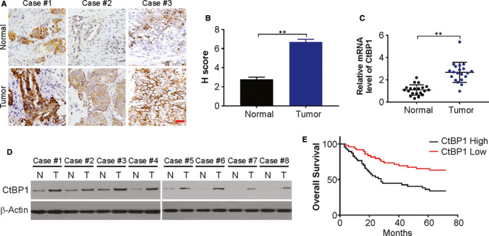
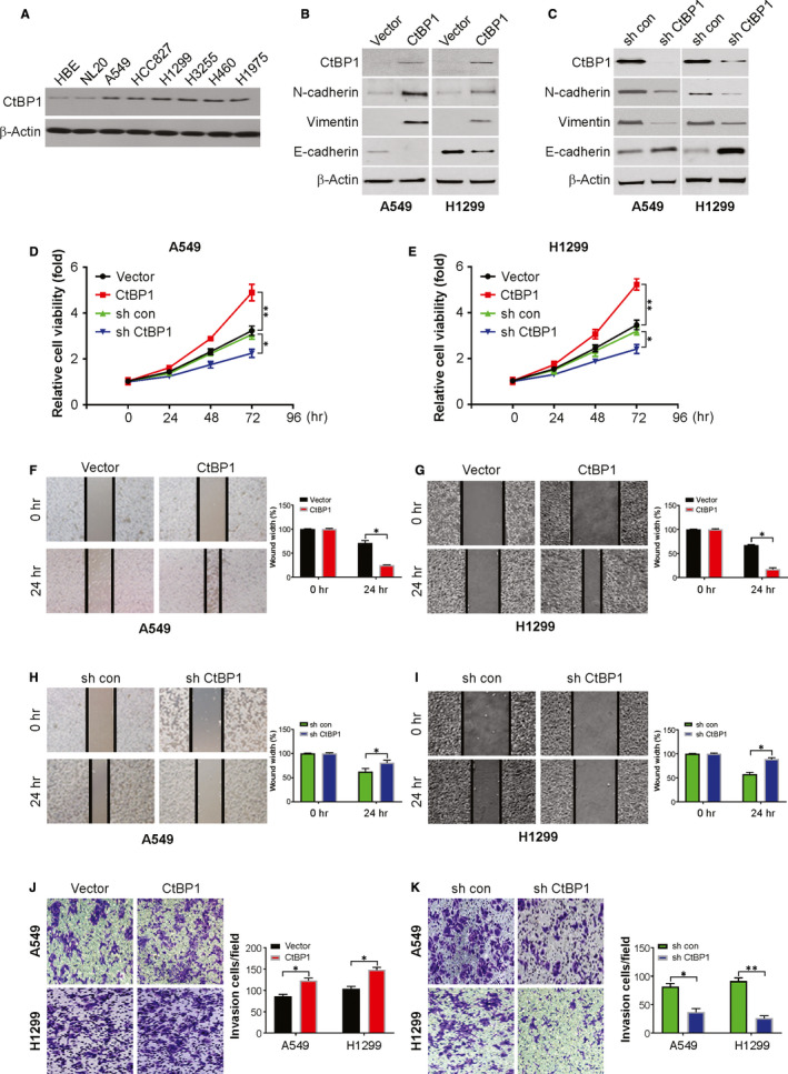
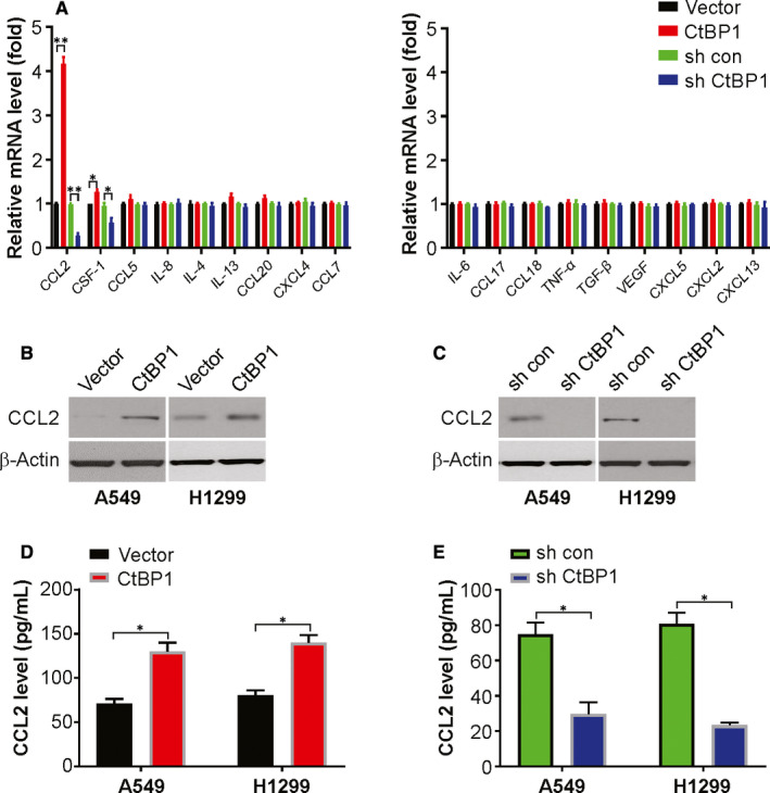
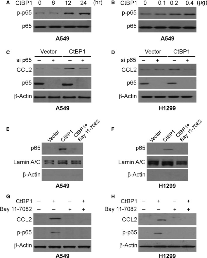
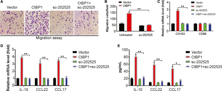
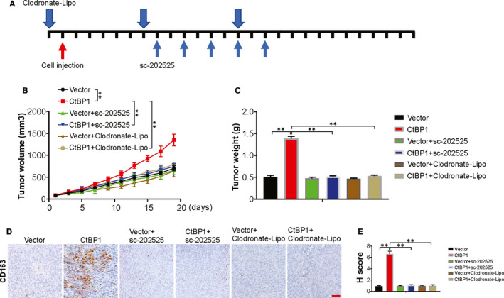
The progression of lung cancer is majorly facilitated by TAMs (tumour-associated macrophages). However, how the TAMs infiltrate the NSCLC microenvironment and the associated biochemical are not fully elaborated. Research has revealed that changes in CtBP1 modulates innate immunity. Here, we investigated if CtBP1 facilitates infiltration of TAM and the subsequent progression of NSCLC. Immunohistochemical analysis was carried out in 96 NSCLC patients to estimate the clinicopathological importance of CtBP1 in the disease. CtBP1 overexpression and knockdown were carried out to assess the activity of CtBP1 in NSCLC cells. Elevated expression of CtBP1 correlated positively with TAMs infiltration into NSCLC tissues, induced EMT (epithelial-mesenchymal transition) in NSCLC cells and modulated the activated NF-κB signalling pathway leading to increase in CCL2 secretion from NSCLC cells, thus promoting TAM recruitment and polarization. TAM induction and polarization reduced significantly on exhausting p65 in NSCLC cells with CtBP1. Moreover, infiltration of TMAs was reduced remarkably on antagonist-mediated blocking of CCR2 and impeded the progression of NSCLC in a mouse model. These findings thus show a novel insight into the process of CtBP1-regulated TAM infiltration in NSCLC.

Keywords: CCL2; CtBP1; NF-κB; NSCLC; TAMs.


Conflict of interest statement

The authors declare that they have no conflict of interest.

Figures

FIGURE 1
FIGURE 1
The expression and clinical significance of CtBP1 in NSCLC. (A) Representative images of IHC staining of CtBP1 expression in 96 human NSCLC tissues and pair‐matched normal tissues. Scale bar, 25 μm. (B) H score of IHC staining of NSCLC tissues and pair‐matched normal tissues. (C) Relative mRNA level of CtBP1 in NSCLC samples and pair‐matched normal tissues was analysed by real‐time PCR. (D) Protein level of CtBP1 in NSCLC samples and pair‐matched normal tissues was analysed by Western blotting. (E) Kaplan‐Meier analysis showing the correlations between CtBP1 expression and the overall survival of patients with NSCLC, determined by log‐rank test. CtBP1 low, n = 48; CtBP1 high, n = 48 (P = 0.023). Data are derived from three independent experiments and presented as mean ± SD. **P < 0.01
FIGURE 2
FIGURE 2
CtBP1 regulates the proliferation, migration and invasion of NSCLC cells in vitro. (A) CtBP1 level in indicated cell lines was analysed by Western blotting. (B) Indicated protein level in control or CtBP1 overexpression cells was analysed by Western blotting. (C) Indicated protein level in sh con or sh CtBP1 cells was analysed by Western blotting. (D) Cell viability of indicated A549 cells was analysed by CCK‐8. (E) Cell viability of indicated H1299 cells was analysed by CCK‐8. (F) Migration of indicated cells was analysed by wound healing assay. (G) Migration of indicated cells was analysed by wound healing assay. (H) Migration of indicated cells was analysed by wound healing assay. (I) Migration of indicated cells was analysed by wound healing assay. (J) Transwell assays were performed to measure the invasion ability of NSCLC cells. (K) Transwell assays were performed to measure the invasion ability of NSCLC cells. Data are derived from three independent experiments and presented as mean ± SD. *P < 0.05, **P < 0.01
FIGURE 3
FIGURE 3
CtBP1 promotes CCL2 induction. (A) Relative mRNA level of indicated cytokines in CtBP1 overexpression or knockdown A549 cells was analysed by real‐time PCR. (B) Protein level of CCL2 in CtBP1 overexpression cells was analysed by Western blotting. (C) Protein level of CCL2 in CtBP1 knockdown cells was analysed by Western blotting. (D) Protein level of CCL2 in CtBP1 overexpression cells was analysed by ELISA. (E) Protein level of CCL2 in CtBP1 knockdown cells was analysed by ELISA. Data are derived from three independent experiments and presented as mean ± SD. *P < 0.05, **P < 0.01
FIGURE 4
FIGURE 4
p65 activation is required for CtBP1‐induced CCL2. (A) A549 cells were transfected with 0.4μg CtBP1. Protein was collected at indicated time points. Expression of p‐p65 (S536) and β‐actin was analysed by Western blotting. (B) A549 cells were transfected with CtBP1 at indicated concentration for 24 hours. Expression of p‐p65 (S536) and β‐actin was analysed by Western blotting. (C) A549 cells were transfected with either a control scrambled siRNA or a p65 siRNA with or without CtBP1 cotransfection for 24 hours. CCL2 expression was analysed by Western blotting. (D) H1299 cells were transfected with either a control scrambled siRNA or a p65 siRNA with or without CtBP1 cotransfection for 24 hours. CCL2 expression was analysed by Western blotting. (E) A549 cells were treated with 10 μmol/L BAY11‐7082 for 1 hour and then transfected with CtBP1 for 24 hours. Nuclear fractions were isolated from cells and analysed for p65 expression by Western blotting. Lamin A/C and β‐actin, which are expressed in nucleus and cytoplasm, respectively, were used as controls for loading and fractionation. (F) H1299 cells were treated with 10 μmol/L BAY11‐7082 for 1 hour and then transfected with CtBP1 for 24 hours. Nuclear fractions were isolated from cells and analysed for p65 expression by Western blotting. Lamin A/C and β‐actin, which are expressed in nucleus and cytoplasm, respectively, were used as controls for loading and fractionation. (G) A549 cells were treated with 10 μmol/L BAY11‐7082 for 1 hour and then transfected with CtBP1 for 24 hours. The levels of p‐p65 (S536) and CCL2 were analysed by Western blotting. (H) H1299 cells were treated with 10 μmol/L BAY11‐7082 for 1 hour, and then transfected with CtBP1 for 24 hours. The levels of p‐p65 (S536) and CCL2 were analysed by Western blotting
FIGURE 5
FIGURE 5
CtBP1 promotes macrophage recruitment and polarization in NSCLC by chemokine (C‐C motif) ligand 2 (CCL2). (A) and (B) Transwell migration assay of macrophage by CM from NSCLC cells as indicated. (C) Real‐time PCR analysis for the expression levels of CD68 and CD163 in THP‐1 macrophages treated with CM from NSCLC cells as indicated. (D) Real‐time PCR for the mRNA expression of tumour‐associated macrophage (TAM) characteristic cytokines in THP‐1 macrophages treated with CM from A549 cells as indicated. (E) Enzyme‐linked immunosorbent assay for the secretion of tumour‐associated macrophage (TAM) characteristic cytokines in THP‐1 macrophages treated with CM from A549 cells as indicated. Data are derived from three independent experiments and presented as mean ± SD. *P < 0.05, **P < 0.01
FIGURE 6
FIGURE 6
CCL2‐mediated TAM recruitment is required for CtBP1‐induced tumour growth. (A) A schematic view of the treatment plan. (B) Tumour volume of indicated tumours. (C) Tumour weight of indicated tumours. (D) IHC staining of CD163 in indicated tumours. Scale bar, 25 μm. (E) H score of IHC staining of CD163 in tumour tissues. Data are derived from three independent experiments and presented as mean ± SD. **P < 0.01

References

    1. Jones GS, Baldwin DR. Recent advances in the management of lung cancer. Clin Med (Lond). 2018;18:s41‐s46. - PMC - PubMed
    1. Shojaee S, Nana‐Sinkam P. Recent advances in the management of non‐small cell lung cancer. F1000Res. 2017;6:2110. - PMC - PubMed
    1. Duma N, Santana‐Davila R, Molina JR. Non‐small cell lung cancer: epidemiology, screening, diagnosis, and treatment. Mayo Clin Proc. 2019;94:1623‐1640. - PubMed
    1. Halliday PR, Blakely CM, Bivona TG. Emerging targeted therapies for the treatment of non‐small cell lung cancer. Curr Oncol Rep. 2019;21:21. - PubMed
    1. Zhang C, Leighl NB, Wu YL, Zhong WZ. Emerging therapies for non‐small cell lung cancer. J Hematol Oncol. 2019;12:45. - PMC - PubMed

Publication types

MeSH terms