. 2020 Nov;177(22):5195-5207.
doi: 10.1111/bph.15258. Epub 2020 Oct 19.

Inhibition of γ-glutamyltransferase ameliorates ischaemia-reoxygenation tissue damage in rats with hepatic steatosis

Affiliations

Inhibition of γ-glutamyltransferase ameliorates ischaemia-reoxygenation tissue damage in rats with hepatic steatosis

Ryuichi Kubota et al. Br J Pharmacol. 2020 Nov.

Abstract

Background and purpose: Hepatic steatosis may be associated with an increased γ-glutamyltransferase (γ-GT) levels. Ischaemia-reoxygenation (IR) injury causes several deleterious effects. We evaluated the protective effects of a selective inhibitor of γ-GT in experimentally induced IR injury in rats with obesity and steatosis.

Experimental approach: Otsuka Long-Evans Tokushima Fatty (OLETF) rats with hepatic steatosis were used in the current study. The portal vein and hepatic artery of left lateral and median lobes were clamped to induce ischaemia. Before clamping, 1 ml of saline (IR group) or 1-ml saline containing 1 mg·kg-1 body weight of GGsTop (γ-GT inhibitor; IR-GGsTop group) was injected into the liver via the inferior vena cava. Blood flow was restored after at 30 min of the start of ischaemia. Blood was collected before, at 30 min after ischaemia and at 2 h and 6 h after reoxygenation. All the animals were killed at 6 h and the livers were collected.

Key results: Treatment with GGsTop resulted in significant reduction of serum ALT, AST and γ-GT levels and hepatic γ-GT, malondialdehyde, 4-hydroxy-2-nonenal and HMGB1 at 6 h after reoxygenation. Inhibition of γ-GT retained normal hepatic glutathione levels. There was prominent hepatic necrosis in IR group, which is significantly reduced in IR-GGsTop group.

Conclusion and implications: Treatment with GGsTop significantly increased hepatic glutathione content, reduced hepatic MDA, 4-HNE and HMGB1 levels and, remarkably, ameliorated hepatic necrosis after ischaemia-reoxygenation. The results indicated that GGsTop could be an appropriate therapeutic agent to reduce IR-induced liver injury in obesity and steatosis.

Keywords: GGsTop; ischaemia; ischaemia-reoxygenation injury; steatosis; γ-glutamyl transpeptidase.


Conflict of interest statement

The authors declare no conflicts of interest.

Figures

FIGURE 1
FIGURE 1
Schematic presentation of the experimental design of ischaemia‐reoxygenation (IR) and GGsTop treatment in rats with hepatic steatosis
FIGURE 2
FIGURE 2
Serum levels of alanine transaminase (ALT), aspartate transaminase (AST) and γ‐glutamyltransferase (γ‐GT) before and after ischaemia‐reoxygenation and effects of GGsTop treatment in rats with hepatic steatosis. (a) Serum levels of ALT. The mean serum ALT level at 6 h after ischaemia‐reoxygenation was significantly lower in IR‐GGsTop group compared to ischaemia‐reoxygenation (IR) group. (b) Serum levels of AST. The mean serum AST level at 6 h after ischaemia‐reoxygenation was significantly less in IR‐GGsTop group compared to IR group. (c) Serum levels of γ‐GT. Serum γ‐GT levels at 2 and 6 h after ischaemia‐reoxygenation maintained same level as sham in GGsTop treated group compared to IR group. The values are mean ± SE (n = 6). **P < 0.01 and ***P < 0.001, when compared to the respective mean values in IR group
FIGURE 3
FIGURE 3
Levels of γ‐glutamyltransferase(γ‐GT), glutathione (GSH) and malondialdehyde (MDA) in the liver tissue of sham, sham‐GGsTop, ischaemia‐reoxygenation (IR) and IR‐GGsTop groups in rats with hepatic steatosis. (a) Hepatic γ‐GT activities. Treatment with GGsTop inhibited the increase of hepatic γ‐GT activity. (b) Hepatic GSH contents. Treatment with GGsTop restored significantly reduced hepatic GSH content in IR group. (c) Hepatic MDA levels. Treatment with GGsTop significantly reduced the elevated MDA levels in IR group. The values are mean ± SE (n = 6)
FIGURE 4
FIGURE 4
Immunohistochemical staining for 4‐hydroxy‐2‐nonenal (4‐HNE) after ischaemia‐reoxygenation and effects of GGsTop treatment in rats with hepatic steatosis. (a) Staining for 4‐HNE in the liver sections from sham, sham‐GGsTop, ischaemia‐reoxygenation (IR) and IR‐GGsTop groups. Moderate staining for 4‐HNE was present in the liver sections from sham and sham‐GGsTop groups in areas with fat globules and surrounding central veins. Marked and strong staining of 4‐HNE was present in pericentral areas and necrotic zone in IR group. However, staining for 4‐HNE was markedly and significantly reduced in IR‐GGsTop group (×40). (b) Quantification of the staining intensity of 4‐HNE. The data are mean ± SE (n = 6)
FIGURE 5
FIGURE 5
Immunohistochemical staining of high mobility group box 1 (HMGB1) after ischaemia‐reoxygenation and effects of GGsTop treatment in rats with hepatic steatosis. (a) Staining for HMGB1 in the liver sections from sham, sham‐GGsTop, ischaemia‐reoxygenation (IR) and IR‐GGsTop groups. Staining for HMGB1 was completely absent in sham and sham‐GGsTop groups. Conspicuous staining of HMGB1 was present in the necrotic zone with fat globules in IR group, while only few cells stained for HMGB1 in IR‐GGsTop group (×100). Inset: higher magnification demonstrating translocation of HMGB1 from nucleus to cytoplasm during ischaemia‐reoxygenation injury (×200). (b) Measurement of the staining area of HMGB1. The data are mean ± SE of six rats per group
FIGURE 6
FIGURE 6
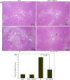
Histopathological alterations in the liver tissue after ischaemia‐reoxygenation and effects of GGsTop treatment in rats with hepatic steatosis. (a) Haematoxylin and eosin (H&E) staining of liver sections from sham, sham‐GGsTop, ischaemia‐reoxygenation (IR) and IR‐GGsTop groups. Fatty degeneration, macrovesicular steatosis, hepatocyte ballooning and moderate necrosis were present especially in pericentral areas in sham and sham‐GGsTop groups. Massive necrosis, sinusoidal congestion and vacuolization of hepatocytes were observed in IR group, which were prominent in pericentral areas. However, only mild congestion and slight necrosis were present in IR‐GGsTop group. Treatment with GGsTop prevented intense hepatic necrosis and retained normal architecture of the liver after reoxygenation (×40). (b) Suzuki's modified IR score. The degree of hepatic IR injury was quantified following Suzuki's criteria based on congestion, vacuolization and necrosis. The data are mean ± SE (n = 6)

References

    1. Ahmadvand, H. , Babaeenezhad, E. , Nasri, M. , Jafaripour, L. , & Khorramabadi, R. M. (2019). Glutathione ameliorates liver markers, oxidative stress and inflammatory indices in rats with renal ischemia reperfusion injury. Journal of Renal Injury Prevention, 8(2), 91–97. 10.15171/jrip.2019.18 - DOI
    1. Alexander, S. P. H. , Roberts, R. E. , Broughton, B. R. S. , Sobey, C. G. , George, C. H. , Stanford, S. C. , … Ahluwalia, A. (2018). Goals and practicalities of immunoblotting and immunohistochemistry: A guide for submission to the British Journal of Pharmacology . British Journal of Pharmacology, 175, 407–411. 10.1111/bph.14112 - DOI - PMC - PubMed
    1. Alexander, S. P. H. , Fabbro, D. , Kelly, E. , Mathie, A. , Peters, J. A. , Veale, E. L. , … Davis, J. A. (2019). The Concise Guide to PHARMACOLOGY 2019/20: Enzymes. British Journal of Pharmacology, 176(Suppl 1), S297–S396. 10.1111/bph.14752 - DOI - PMC - PubMed
    1. Ayala, A. , Muñoz, M. F. , & Argüelles, S. (2014). Lipid peroxidation: Production, metabolism, and signaling mechanisms of malondialdehyde and 4‐hydroxy‐2‐nonenal. Oxidative Medicine and Cellular Longevity, 2014, 360438 10.1155/2014/360438 PMID: 24999379 - DOI - PMC - PubMed
    1. Behrns, K. E. , Tsiotos, G. G. , DeSouza, N. F. , Krishna, M. K. , Ludwig, J. , & Nagorney, D. M. (1998). Hepatic steatosis as a potential risk factor for major hepatic resection. Journal of Gastrointestinal Surgery, 2(3), 292–298. 10.1016/s1091-255x(98)80025-5 PMID: 9841987 - DOI - PubMed

Publication types