

Peripapillary Retinal Vascular Involvement in Early Post-COVID-19 Patients
- PMID: 32911619
- PMCID: PMC7565672
- DOI: 10.3390/jcm9092895
Peripapillary Retinal Vascular Involvement in Early Post-COVID-19 Patients
Abstract
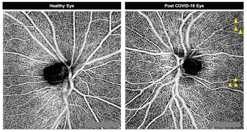
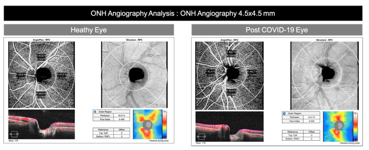
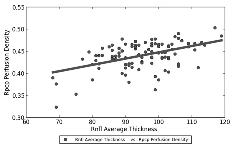
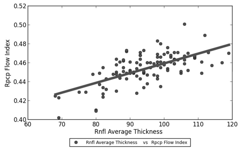
The ability of severe acute respiratory syndrome coronavirus 2 (SARS-CoV-2's) to cause multi-organ ischemia and coronavirus-induced posterior segment eye diseases in mammals gave concern about potential sight-threatening ischemia in post coronavirus disease 2019 patients. The radial peripapillary capillary plexus (RPCP) is a sensitive target due to the important role in the vascular supply of the peripapillary retinal nerve fiber layer (RNFL). Eighty patients one month after SARS-CoV-2 infection and 30 healthy patients were selected to undergo structural OCT (optical coherence tomography) and OCTA (optical coherence tomography angiography) exams. Primary outcome was a difference in RPCP perfusion density (RPCP-PD) and RPCP flow index (RPCP-FI). No significant difference was observed in age, sex, intraocular pressure (IOP) and prevalence of myopia. RPCP-PD was lower in post SARS-CoV-2 patients compared to controls. Within the post-COVID-19 group, patients with systemic arterial hypertension had lower RPCP-FI and age was inversely correlated to both RPCP-FI and RPCP-PD. Patients treated with lopinavir + ritonavir or antiplatelet therapy during admission had lower RPCP-FI and RPCP-PD. RNFL average thickness was linearly correlated to RPCP-FI and RPCP-PD within post-COVID-19 group. Future studies will be needed to address the hypothesis of a microvascular retinal impairment in individuals who recovered from SARS-CoV-2 infection.
Keywords: OCT angiography; SARS-CoV-2; peripapillary capillary perfusion; personalized medicine.
Conflict of interest statement
No conflicting relationship exists for any author.
Figures
References
-
- WHO . Summary Table of SARS Cases by Country N–A. World Health Organisation; Geneva, Switzerland: Nov 1, 2002.
-
- WHO . Organisation WH Coronavirus Disease 2019 (COVID-19) World Health Organisation; Geneva, Switzerland: Mar, 2020. Situation Report 32.
LinkOut - more resources
Full Text Sources
Miscellaneous
