

A review of virtual planning software for guided implant surgery - data import and visualization, drill guide design and manufacturing
- PMID: 32912273
- PMCID: PMC7488021
- DOI: 10.1186/s12903-020-01208-1
A review of virtual planning software for guided implant surgery - data import and visualization, drill guide design and manufacturing
Abstract
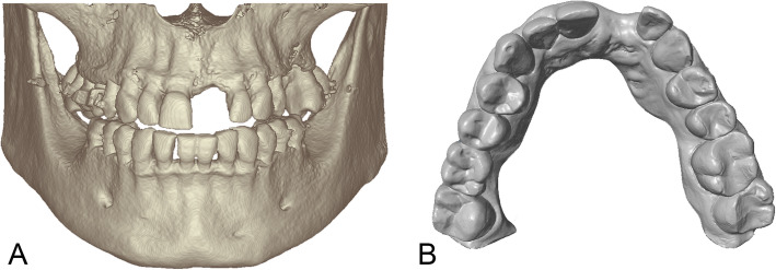
Background: Virtual implant planning systems integrate (cone beam-) computed tomography data to assess bone quantity and virtual models for the design of the implant-retained prosthesis and drill guides. Five commercially available systems for virtual implant planning were examined regarding the modalities of integration of radiographic data, virtual dental models and the design of drill guides for guided implant surgery. The purpose of this review was to describe the limitations of these available systems regarding the import of imaging data and the design and fabrication of a drill guide.
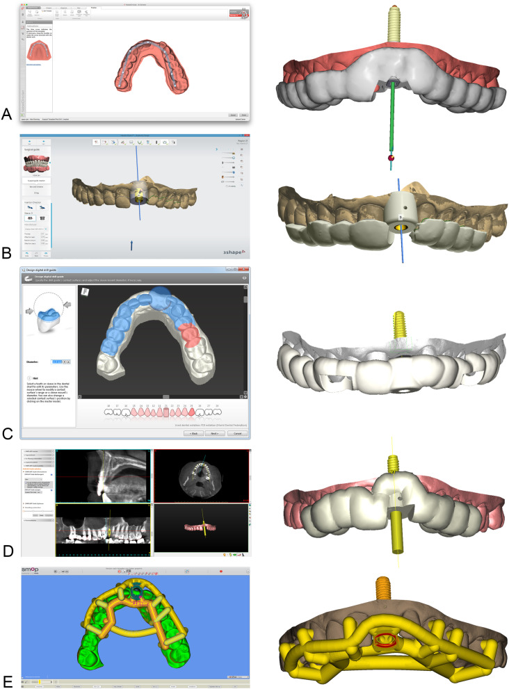
Methods: The following software systems were examined regarding the import of imaging data and the export of the virtual implant planning for the design and fabrication of a drill guide with the help of two clinical situations requiring dental implant therapy: coDiagnostiX™, DentalWings, Canada (CDX); Simplant Pro™, Dentsply, Sweden (SIM); Smop™, Swissmeda, Switzerland (SMP); NobelClinician™, Nobel Biocare, Switzerland (NC); Implant Studio, 3Shape, Denmark (IST). Assessment criteria included data formats and management as well as the workflow for the design and production of drill guides.
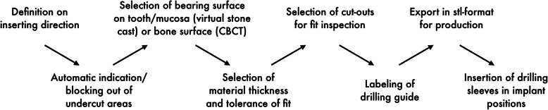
Results: All systems have a DICOM-interface ("Digital Imaging and Communication in Medicine") for the import of radiographic data. Imaging artefacts could be reduced but not eliminated by manual data processing. The import of virtual dental models in a universal format (STL: Standard Tesselation Language) was possible with three systems; one system could only be used with a proprietary data format. All systems display three-dimensional surface models or two-dimensional cross-sections with varying orientation for virtual implant planning. Computer aided design and manufacturing (CAD/CAM) of drill guides may be performed by the user with the help of default parameters or solely by the provider of the software and thus without the influence of the clinician.
Conclusion: Data bases of commonly used implant systems are available in all tested software, however not all systems allow to plan and execute fully guided implant placement. An individual design and in-house manufacturing of the drill guide is only available in some software systems. However, at the time of publication most recent software versions showed flexibility in individual design and in-house manufacturing of drill guides.
Keywords: Computer-aided design; Computer-assisted surgery; Guided implant surgery; Virtual implant planning.
Conflict of interest statement
The authors declare that they have no competing interests.
Figures
References
Publication types
MeSH terms
Substances
LinkOut - more resources
Full Text Sources
Other Literature Sources
Miscellaneous
