Case Reports
. 2020 Sep 9;13(9):e234830.
doi: 10.1136/bcr-2020-234830.

Primary malignant melanoma of the stomach: a rare entity

Affiliations
Case Reports

Primary malignant melanoma of the stomach: a rare entity

Pradeep Zechariah et al. BMJ Case Rep. .

Abstract

A 54-year-old man presented with easy fatiguability, dyspnoea on exertion and dyspeptic symptoms. On evaluation, he was found to have an ulcero-proliferative growth in the gastric fundus, the biopsy of which was malignant melanoma of the stomach. Further evaluation with 18F-fluorodeoxyglucose positron emission tomography-computed tomography (18F-FDG PET-CT) scan showed operable disease with no focus of disease elsewhere. He was diagnosed as primary gastric melanoma and underwent radical total gastrectomy with adequate margins. His postoperative period was uneventful. Further adjuvant therapy was refused by the patient. At 6-month follow-up, an 18F-FDG PET-CT scan was done, which showed no evidence of disease. On follow-up at 1-year, he was alive and asymptomatic.

Keywords: gastric cancer; gastrointestinal surgery; malignant disease and immunosuppression; pathology; stomach and duodenum.


Conflict of interest statement

Competing interests: None declared.

Figures

Figure 1
Figure 1
Upper gastrointestinal endoscopy showing the ulceroproliferative growth in the gastric fundus, extending to the cardia.
Figure 2
Figure 2
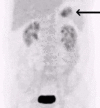
Positron emission tomography-computed tomography scan of the abdomen on admission showing increased tracer activity in the stomach (black arrow).
Figure 3
Figure 3
Contrast-enhanced computed tomography scan of the abdomen done as part of the PET-CT showing the gastric fundus growth (blue arrow).
Figure 4
Figure 4
Positron emission tomography-computed tomography scan of the abdomen showing metabolically active focus in the gastric fundus (black arrow).
Figure 5
Figure 5
Gross surgical specimen showing the ulcero-proliferative growth in the gastric fundus (yellow arrow).
Figure 6
Figure 6
Magnified view of the gastric fundal growth showing the central deep ulceration (yellow arrow).
Figure 7
Figure 7
H&E staining (40X).
Figure 8
Figure 8
H&E staining (100X).
Figure 9
Figure 9
H&E staining (200X).
Figure 10
Figure 10
Photomicrograph showing immunohistochemistry positivity for Melan-A.
Figure 11
Figure 11
Photomicrograph showing immunohistochemistry positivity for HMB-45.
Figure 12
Figure 12
Positron emission tomography-computed tomography (PET-CT) scan on follow-up.

References

    1. Ali Z, Yousaf N, Larkin J. Melanoma epidemiology, biology and prognosis. EJC Suppl 2013;11:81–91. 10.1016/j.ejcsup.2013.07.012 - DOI - PMC - PubMed
    1. Bramhall RJ, Mahady K, Peach AHS. Spontaneous regression of metastatic melanoma - clinical evidence of the abscopal effect. Eur J Surg Oncol 2014;40:34–41. 10.1016/j.ejso.2013.09.026 - DOI - PubMed
    1. Paolino G, Didona D, Macrì G, et al. . Anorectal Melanoma : Scott JF, Gerstenblith MR, Noncutaneous Melanoma [Internet. Brisbane (AU: Codon Publications, 2018. https://www.ncbi.nlm.nih.gov/books/NBK506984/ - PubMed
    1. Callaghan GM, Kelleher FC, Ridgway PF, et al. . A Case of Primary Gastric Melanoma Exhibiting a Rare BRAF V600R Mutation. Eur J Case Rep Intern Med 2018;5:1. 10.12890/2018_000749 - DOI - PMC - PubMed
    1. Wiewiora M, Steplewska K, Piecuch JZ, et al. . A rare case of primary gastric melanoma. Indian J Surg 2019.

Publication types

MeSH terms

Substances