Multicenter Study
. 2020 Nov 24;95(21):e2866-e2879.
doi: 10.1212/WNL.0000000000010794. Epub 2020 Sep 10.

Cardiac phenotype in ATP1A3-related syndromes: A multicenter cohort study

Affiliations
Multicenter Study

Cardiac phenotype in ATP1A3-related syndromes: A multicenter cohort study

Simona Balestrini et al. Neurology. .

Abstract

Objective: To define the risks and consequences of cardiac abnormalities in ATP1A3-related syndromes.

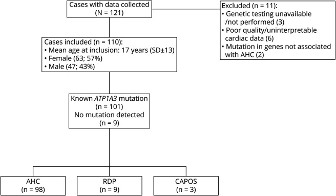
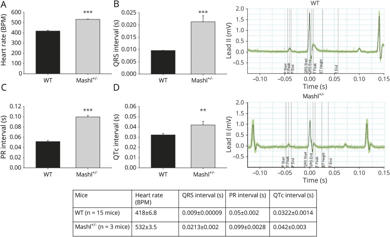
Methods: Patients meeting clinical diagnostic criteria for rapid-onset dystonia-parkinsonism (RDP), alternating hemiplegia of childhood (AHC), and cerebellar ataxia, areflexia, pes cavus, optic atrophy, and sensorineural hearing loss (CAPOS) with ATP1A3 genetic analysis and at least 1 cardiac assessment were included. We evaluated the cardiac phenotype in an Atp1a3 knock-in mouse (Mashl+/-) to determine the sequence of events in seizure-related cardiac death.

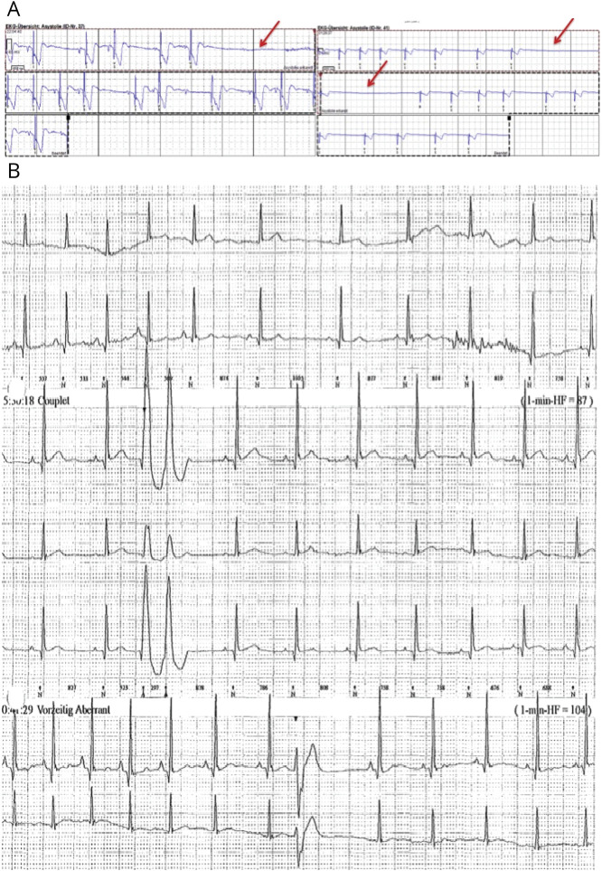
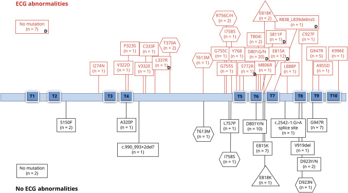
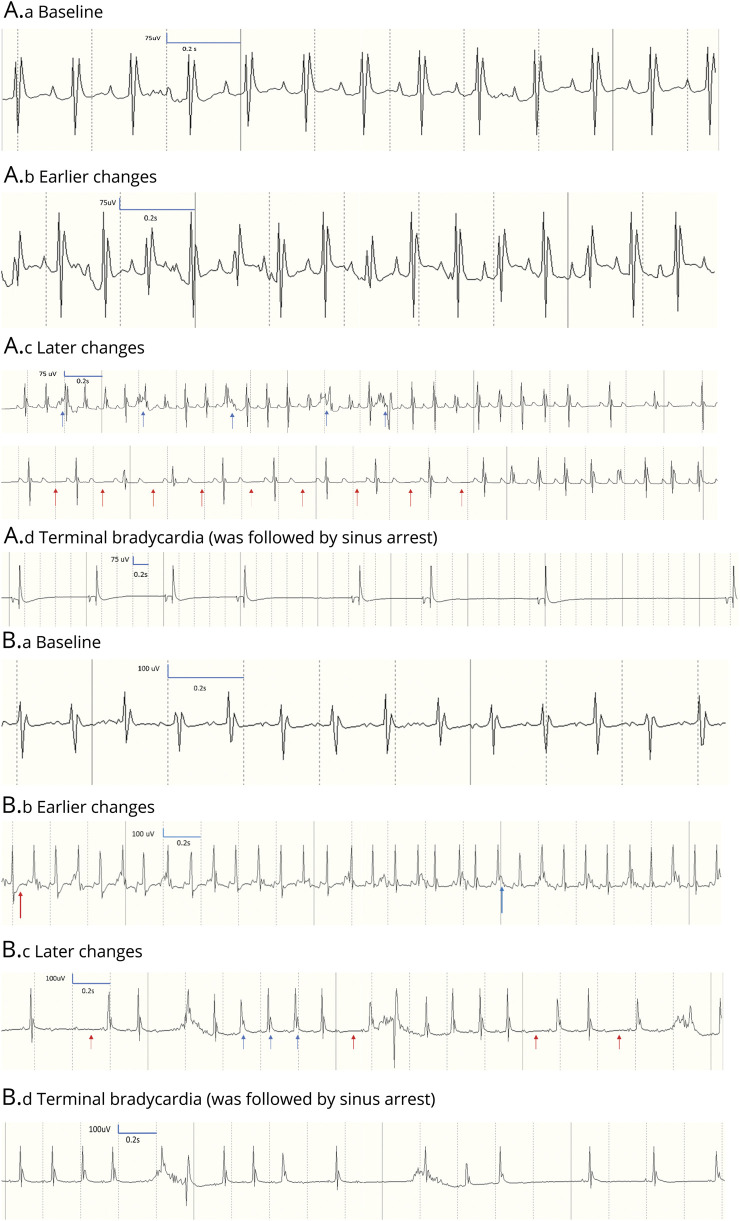
Results: Ninety-eight patients with AHC, 9 with RDP, and 3 with CAPOS (63 female, mean age 17 years) were included. Resting ECG abnormalities were found in 52 of 87 (60%) with AHC, 2 of 3 (67%) with CAPOS, and 6 of 9 (67%) with RDP. Serial ECGs showed dynamic changes in 10 of 18 patients with AHC. The first Holter ECG was abnormal in 24 of 65 (37%) cases with AHC and RDP with either repolarization or conduction abnormalities. Echocardiography was normal. Cardiac intervention was required in 3 of 98 (≈3%) patients with AHC. In the mouse model, resting ECGs showed intracardiac conduction delay; during induced seizures, heart block or complete sinus arrest led to death.

Conclusions: We found increased prevalence of ECG dynamic abnormalities in all ATP1A3-related syndromes, with a risk of life-threatening cardiac rhythm abnormalities equivalent to that in established cardiac channelopathies (≈3%). Sudden cardiac death due to conduction abnormality emerged as a seizure-related outcome in murine Atp1a3-related disease. ATP1A3-related syndromes are cardiac diseases and neurologic diseases. We provide guidance to identify patients potentially at higher risk of sudden cardiac death who may benefit from insertion of a pacemaker or implantable cardioverter-defibrillator.


Figures

Figure 1
Figure 1. Study design
Diagram illustrating study design with included and excluded cases. AHC = alternating hemiplegia of childhood; CAPOS = cerebellar ataxia, areflexia, pes cavus, optic atrophy, and sensorineural hearing loss; RDP = rapid-onset dystonia-parkinsonism.
Figure 2
Figure 2. Holter abnormalities in a young patient with AHC requiring the insertion of an ICD
(A) Abnormal Holter ECG showing asymptomatic sinus pauses of up to 4 seconds in duration (red arrows) and (B) polymorphic ventricular ectopics in couplets and bigeminy in a young patient who required insertion of an implantable cardioverter-defibrillator (ICD) at the age of 27 years (patient with alternating hemiplegia of childhood [AHC], c.2401G>A p.D801N mutation).
Figure 3
Figure 3. ECG abnormalities in ATP1A3-related syndromes
Graphic representation of the ATP1A3-related syndromes (alternating hemiplegia of childhood [AHC] = rectangles, cerebellar ataxia, areflexia, pes cavus, optic atrophy, and sensorineural hearing loss [CAPOS] = triangles, rapid-onset dystonia-parkinsonism [RPD] = hexagons) (n = 110), associated mutations, and prevalence of ECG (12-lead and/or Holter) abnormalities. Each box includes the number of cases with a specific mutation and ECG abnormalities in the upper part or without ECG abnormalities in the lower part. Reference sequences for the corresponding ATP1A3 transcript and protein were NM_152296.4 and Uniprot P13637, respectively. T1 through T10 are transmembrane domains. The position of the mutation across the functional domains was not associated with different prevalence of ECG abnormalities or dynamic changes. In 9 patients with AHC, no mutation was identified in ATP1A3. D = dynamic changes (when serial tests were available).
Figure 4
Figure 4. ATP1A3 expression in an adult normal heart
Dark brown stripes show strong immunolabeling for ATP1A3 corresponding to intercalated disks in adult myocardium from a 75-year-old man (cause of death at postmortem: bronchopneumonia). Tissue samples were fixed in formalin and embedded in paraffin. A standard immunohistochemistry method was applied to 5-μm-thick sections with primary antibody anti-ATP1A3 (Santa Cruz, polyclonal, goat, sc16052) at a dilution of 1:1,000 with overnight incubation at 4°C in diluent buffer (DAKO REAL, Ab diluent S2022). Immunostaining was qualitatively evaluated.
Figure 5
Figure 5. ECG data in the Mashl+/− compared to WT mice
Comparison of ECG data acquired from wild-type (WT; n = 15) and Mashl+/− mice (n = 3). (A) Heart rate, (B) QRS interval, (C) PR interval, and (D) QTc interval. Traces are examples of ECG traces in WT and mutant mice. Heart rate, QRS, and PR interval were higher in Mashl+/− mice. ***p ≤ 0.001, **p < 0.01 (Student t test). BPM = beats per minute.
Figure 6
Figure 6. ECG abnormalities in the Mashl+/− mice (n = 3) after seizure induction
(A) Mashl+/− No. 1 ECG traces. (A.a) Baseline, with normal heart rate, noise present stems from skeletal muscle (breathing) activity. (A.b) Earlier changes started with onset of EEG seizures at 21 minutes after injection: heart rate increase and JT-segment elevation. (A.c) Later changes. Consecutive ECG traces shortly before sinus arrest showing heart rate fluctuation and atrioventricular block (red arrows); premature ventricular contractions are also visible (blue arrows). (A.d) Terminal change. Sinus bradycardia that was followed by sinus arrest. (B) Mashl+/− No. 2 ECG traces. (B.a) Baseline, with normal heart rate. (B.b) Earlier changes started with onset of EEG seizures after 5 minutes of injection and persisted to 1 minute before death (time of B.b. illustration). Increased heart rate with JT-segment depression (red), and JT elevation (blue) widened QRS. (B.c) Later changes. Sequence of events <1 minute before sinus arrest showing atrioventricular block (red arrows) and elevated JT segments (blue arrows). (B.d) Terminal change. Sinus bradycardia that was followed by sinus arrest. Mashl+/− No. 3 had similar ECG traces from baseline to terminal changes.

References

    1. Heinzen EL, Swoboda KJ, Hitomi Y, et al. . De novo mutations in ATP1A3 cause alternating hemiplegia of childhood. Nat Genet 2012;44:1030–1034. - PMC - PubMed
    1. de Carvalho Aguiar P, Sweadner KJ, Penniston JT, et al. . Mutations in the Na(+)/K(+)-ATPase alpha-3 gene ATP1A3 are associated with rapid-onset dystonia parkinsonism. Neuron 2004;43:169–175. - PubMed
    1. Demos MK, van Karnebeek CDM, Ross CJD, et al. . A novel recurrent mutation in ATP1A3 causes CAPOS syndrome. Orphanet J Rare Dis 2014;9:15. - PMC - PubMed
    1. Brashear A, Sweadner KJ, Cook JF, Swoboda KJ, Ozelius L. ATP1A3-Related neurologic disorders. 2008. In: Adam MP, Ardinger, HH, Pagon RA, et al.., editors. GeneReviews® [Internet]. Seattle: University of Washington; 1993–2018. Available at: ncbi.nlm.nih.gov/books/NBK1115/. Accessed January 5, 2019. - PubMed
    1. Masoud M, Prange L, Wuchich J, et al. . Diagnosis and treatment of alternating hemiplegia of childhood. Curr Treat Options Neurol 2017;19:8. - PubMed

Publication types

MeSH terms

Substances

Supplementary concepts