. 2020 Sep 11;369(6509):eaba3066.
doi: 10.1126/science.aba3066.

The impact of sex on gene expression across human tissues

Collaborators, Affiliations

The impact of sex on gene expression across human tissues

Meritxell Oliva et al. Science. .

Abstract

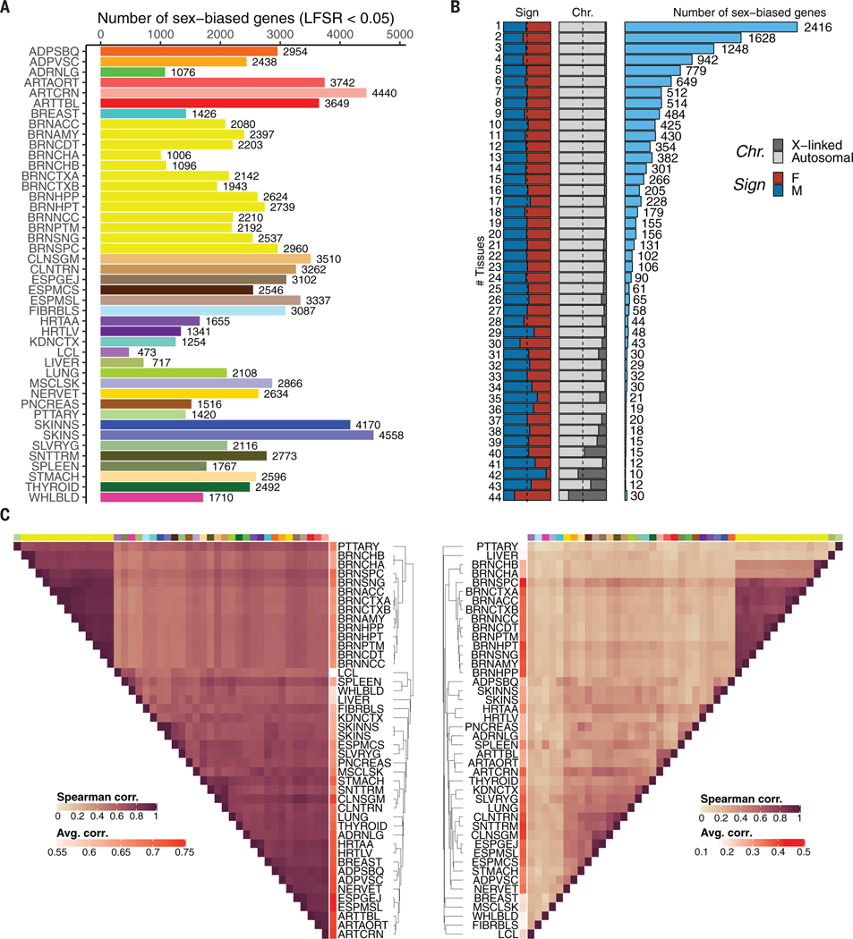
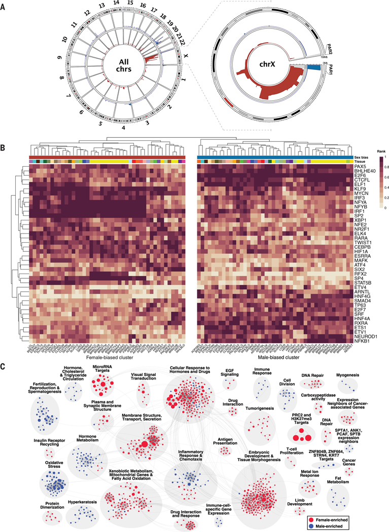
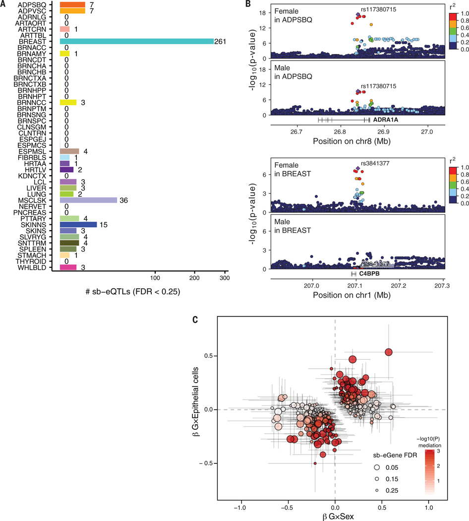
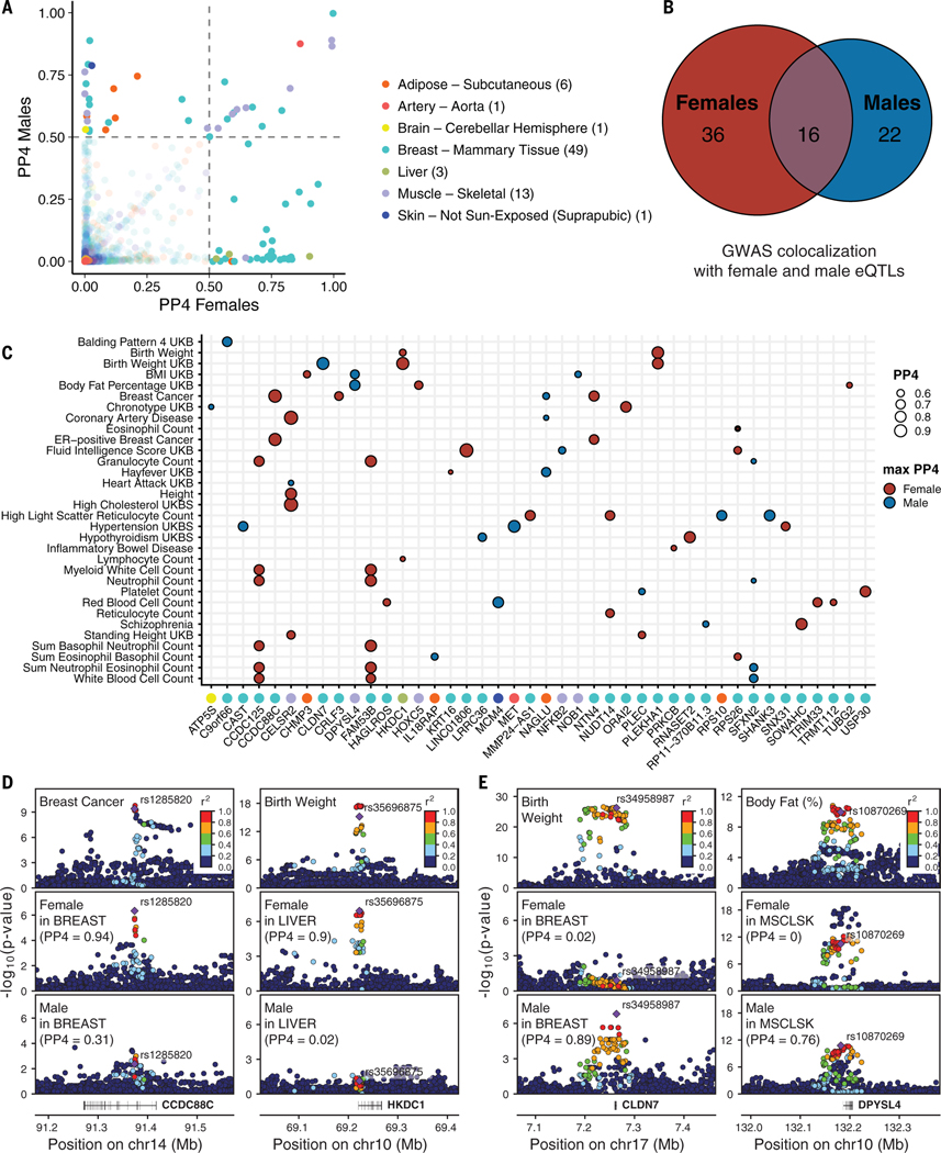
Many complex human phenotypes exhibit sex-differentiated characteristics. However, the molecular mechanisms underlying these differences remain largely unknown. We generated a catalog of sex differences in gene expression and in the genetic regulation of gene expression across 44 human tissue sources surveyed by the Genotype-Tissue Expression project (GTEx, v8 release). We demonstrate that sex influences gene expression levels and cellular composition of tissue samples across the human body. A total of 37% of all genes exhibit sex-biased expression in at least one tissue. We identify cis expression quantitative trait loci (eQTLs) with sex-differentiated effects and characterize their cellular origin. By integrating sex-biased eQTLs with genome-wide association study data, we identify 58 gene-trait associations that are driven by genetic regulation of gene expression in a single sex. These findings provide an extensive characterization of sex differences in the human transcriptome and its genetic regulation.


Figures

Fig. 1.
Fig. 1.. Sample, data types, and discovery sets in the study of sex differences in GTEx v8.
Tissue types (including 11 distinct brain regions and two cell lines) are illustrated, with sample numbers from GTEx v8 genotyped donors (females:males, in parentheses) and color coding indicated for each. This study included N = 44 tissue sources present in both sexes with ≥70 samples. Tissue sources comprised two cell lines, 40 tissues, and two additional replicates for brain cerebellum and cortex tissues. Tissue name abbreviations are shown in bold. See (9) for specific numbers of donors used in each analysis.
Fig. 2.
Fig. 2.. Sex-differential gene expression.
(A) Number of sex-differentially expressed genes (sex-biased genes) per tissue. Tissue colors are as in Fig. 1. (B) Sex-biased gene discovery (histogram, number of sex-biased genes) and characteristics of sex-biased genes (stacked bar plots) as a function of tissue sharing. Proportions of X-linked and autosomal sex-biased genes (Chr.) and of female- and male-biased genes (Sign) are indicated. (C) Hierarchical clustering of tissues based on gene expression (left) and the effect size of sex-biased genes (right). See (9) for further details.
Fig. 3.
Fig. 3.. Regulatory mechanisms and biological functions of sex-biased genes.
(A) Genomic position enrichment of sex-biased genes, as indicated by male-biased (blue) and female-biased (red) genes across all chromosomes (left) and chromosome X (right). The height of each rim represents the tissue sharing of the significant genomic enrichment signal and ranges from 1 to 44 (number of tissue sources). See (9) for further details. (B) Transcription factor binding site (TFBS) enrichment in promoter regions of sex-biased genes. Of 92 enriched TFBS profiles, the top 40 with the largest difference across all tissues in the enrichment profile derived from male-biased and female-biased genes are displayed. Values represent the TFBS enrichment ranking transformed to [0, 1] per tissue and per sex; a value of 1 corresponds to the highest enrichment. See (9) for further details. (C) Clusters (gray circles) of gene sets enriched for genes highly expressed (blue and red balloons) in females (red) or males (blue) across tissues. Balloon size corresponds to the P value for the across-tissue meta-analysis of GSEA. Faint lines connecting balloons correspond to shared leading-edge genes between gene sets. See (9) for further details.
Fig. 4.
Fig. 4.. Sex-biased eQTLs (sb-eQTLs).
(A) Number of sb-eQTLs discovered per tissue. Square-root transformation was applied to the x axis. See Fig. 1 for tissue abbreviations. (B) Association P values of the female-stratified (top) and male-stratified (bottom) cis-eQTLs in the ADRA1A locus in adipose subcutaneous tissue (upper panels; βF = −0.78, PF = 4.64 × 10⁻¹⁸, βM = −0.47, PM = 3.98 × 10⁻¹⁰, PG×Sex = 1.05 × 10⁻⁵) and C4BPB locus in breast mammary tissue (lower panels; βF = 0.40, PF = 2.68 × 10⁻⁷, βM = −0.02, PM = 0.89, PG×Sex = 7.22 × 10⁻⁵). Linkage disequilibrium between loci is quantified by squared Pearson coefficient of correlation (r2). Diamond-shaped point represents the top significant eQTL variant across sex-stratified P values. (C) sb-eQTL mediation analysis of 261 breast sb-eQTLs. Point coordinates represent the effect size of G×Sex (x axis) and G×Epithelial cells (y axis) derived from a linear regression model with both interaction terms. Gray lines represent confidence intervals of the effect sizes of G×Sex (horizontal lines) and G×Epithelial cells (vertical lines). Point size represents sb-eQTL significance; color corresponds to mediation significance. See (9) for further details.
Fig. 5.
Fig. 5.. Colocalization of sb-eQTLs with GWAS traits.
(A) Posterior probability (PP4) of 74 colocalized gene-trait pairs where a GWAS shows evidence of colocalization with the female-stratified and/or male-stratified cis-eQTL signal (PP4 > 0.5). Numbers of colocalizing loci per tissue are shown in parentheses. (B) Numbers of colocalizing loci for female and male cis-eQTLs. (C) GWAS-eQTL colocalizing genes (PP4 > 0.5) color-labeled by eQTL tissue of origin according to labels in (A) (x axis) are categorized by the sex where the colocalization signal is maximized with the corresponding GWAS trait (y axis). Comparing the colocalization PP4 values for male and female cis-eQTL signals, the estimates can be maximum in females (red) or males (blue). (D) Genotype-phenotype association P values of the CCDC88C (left) and HKDC1 (right) loci. For the CCDC88C locus, panels illustrate GWAS signal for breast cancer (top) and CCDC88C cis-eQTL signal for females (middle) and males (bottom) in breast mammary tissue. For the HKDC1 locus, panels illustrate GWAS signal for birth weight (top) and HKDC1 cis-eQTL signal for females (middle) and males (bottom) in liver. (E) Genotype-phenotype association P values of the CLDN7 (left) and DPYSL4 (right) loci. For the CLDN7 locus, panels illustrate GWAS signal for birth weight (top) and CLDN7 cis-eQTL signal for females (middle) and males (bottom) in breast mammary tissue. For the DPYSL4 locus, panels illustrate GWAS signal for body fat (top) and DPYSL4 cis-eQTL signal for females (middle) and males (bottom) in muscle skeletal tissue. In (D) and (E), linkage disequilibrium between loci is quantified by squared Pearson coefficient of correlation (r2). Diamond-shaped point represents the top significant cis-eQTL variant across sex-stratified P values.

Comment in

Similar articles

Cited by

References

    1. Zheng D et al. , Sexual dimorphism in the incidence of human cancers. BMC Cancer 19, 684 (2019). doi: 10.1186/s12885-019-5902-z; pmid: 31299933 - DOI - PMC - PubMed
    1. Anderson GD, Sex and racial differences in pharmacological response: Where is the evidence? Pharmacogenetics, pharmacokinetics, and pharmacodynamics. J. Womens Health 14, 19–29 (2005). doi: 10.1089/jwh.2005.14.19; pmid: 15692274 - DOI - PubMed
    1. Kuan V et al. , A chronological map of 308 physical and mental health conditions from 4 million individuals in the English National Health Service. Lancet Digit. Health 1, e63–e77 (2019). doi: 10.1016/S2589-7500(19)30012-3; pmid: 31650125 - DOI - PMC - PubMed
    1. Ngo ST, Steyn FJ, McCombe PA, Gender differences in autoimmune disease. Front. Neuroendocrinol. 35, 347–369 (2014). doi: 10.1016/j.yfrne.2014.04.004; pmid: 24793874 - DOI - PubMed
    1. Westergaard D, Moseley P, Sørup FKH, Baldi P, Brunak S, Population-wide analysis of differences in disease progression patterns in men and women. Nat. Commun. 10, 666 (2019). doi: 10.1038/s41467-019-08475-9; pmid: 30737381 - DOI - PMC - PubMed

Publication types

Grants and funding