. 2020 Sep 10;11(9):738.
doi: 10.1038/s41419-020-02949-1.

A small molecular compound CC1007 induces cross-lineage differentiation by inhibiting HDAC7 expression and HDAC7/MEF2C interaction in BCR-ABL1- pre-B-ALL

Affiliations

A small molecular compound CC1007 induces cross-lineage differentiation by inhibiting HDAC7 expression and HDAC7/MEF2C interaction in BCR-ABL1- pre-B-ALL

Zhihua Wang et al. Cell Death Dis. .

Abstract

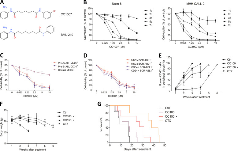
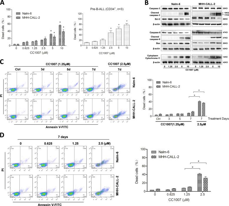
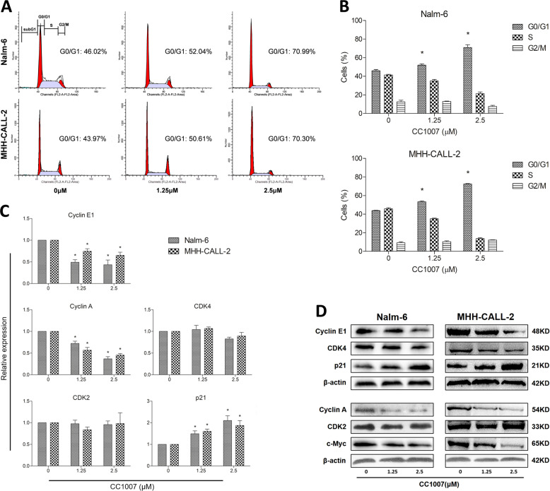
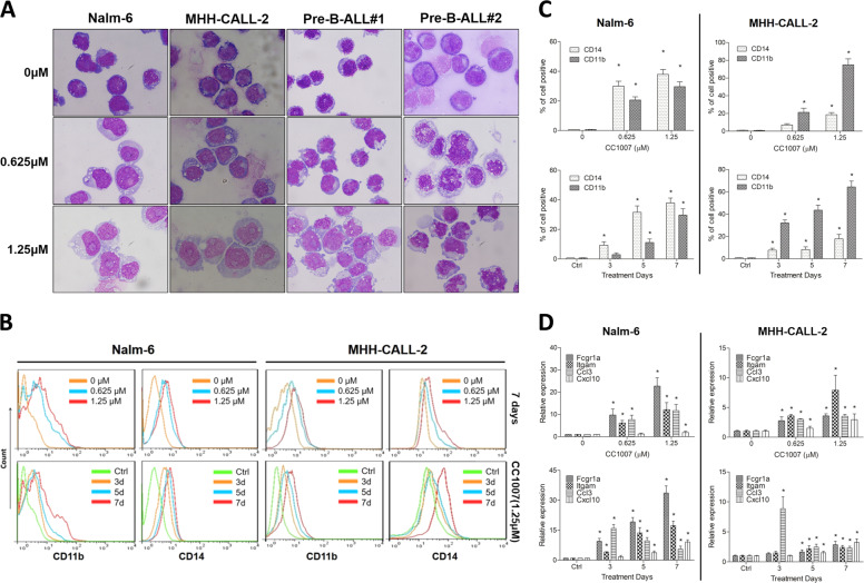
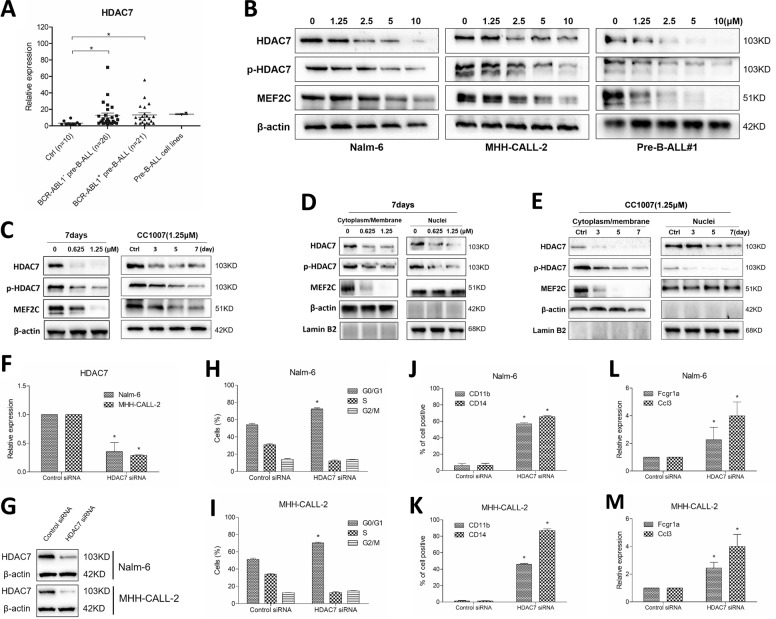
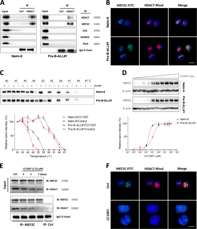
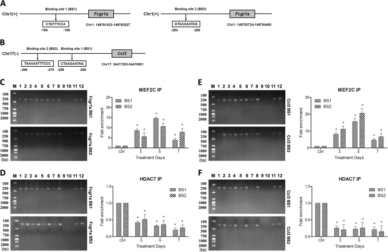
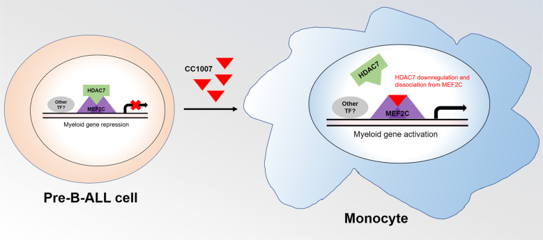
Histone deacetylase 7 (HDAC7), a member of class IIa HDACs, has been described to be an important regulator for B cell development and has a potential role in B cell acute lymphoblastic leukemia (B-ALL). CC1007, a BML-210 analog, is designed to indirectly inhibit class IIa HDACs by binding to myocyte enhancer factor-2 (MEF2) and blocking the recruitment of class IIa HDACs to MEF2-targeted genes to enhance the expression of these targets. In this study, we investigated the anticancer effects of CC1007 in breakpoint cluster region-Abelson 1 fusion gene-negative (BCR-ABL1-) pre-B-ALL cell lines and primary patient-derived BCR-ABL1- pre-B-ALL cells. CC1007 had obvious antileukemic activity toward pre-B-ALL cells in vitro and in vivo; it also significantly prolonged median survival time of pre-B-ALL-bearing mice. Interestingly, low dose of CC1007 could inhibit proliferation of BCR-ABL1- pre-B-ALL cells in a time-dependent manner not accompanied by significant cell apoptosis, but along with cross-lineage differentiation toward monocytic lineage. From a mechanistic angle, we showed that HDAC7 was overexpressed in BCR-ABL1- pre-B-ALL cells compared to normal bone marrow samples, and CC1007 could reduce the binding of HDAC7 at the promoters of monocyte-macrophage-specific genes via inhibition of HDAC7 expression and HDAC7:MEF2C interaction. These data indicated that CC1007 may be a promising agent for the treatment of BCR-ABL1- pre-B-ALL.


Conflict of interest statement

The authors declare that they have no conflict of interest.

Figures

Fig. 1
Fig. 1. Antiproliferative effect of CC1007 in vitro and in vivo.
a Chemical structure of CC1007 and BML-210. b Effects of CC1007 on BCR-ABL1 pre-B-ALL cell line Nalm-6 and MHH-CALL-2 cell growth (MTT assay). The cell growth curve represents the effect of CC1007 at different concentrations for 7 days. The values represent the mean ± SEM of triplicate cultures. c Effects of CC1007 on fresh CD34+ cell growth from BCR-ABL1 pre-B-ALL patients (n = 15), fresh mononuclear cell (MNC) growth from BCR-ABL1 pre-B-ALL patients (n = 20), and fresh MNC growth from normal controls (n = 10). Cells were treated with CC1007 for 72 h, and then cell viability was evaluated by the MTT assay. d Effect of CC1007 on BCR-ABL1+ primary CD34+ cell (n = 8) or MNC (n = 8) growth and BCR-ABL1 primary CD34+ cell (n = 15) or MNC (n = 20) growth from pre-B-ALL patients. Cells were treated with CC1007 for 72 h. The growth curve represents the effect of CC1007 at different concentrations, which are shown as the mean ± SEM. e Examination was performed on human white blood cell (WBC) antigen (CD45) levels to evaluate the effect of CC1007 on the proliferation of human primary BCR-ABL1 pre-B-ALL cells in a xenograft model. The results represent the mean ± SEM. f Changes of body weight of mice treated with CC1007 and CTX. The results represent the mean ± SEM. g A Kaplan–Meier survival plot for BCR-ABL1 pre-B-ALL-bearing NOD/SCID mice is shown. The survival curves differed significantly between the CC1007 (150 mg/kg)-treated group and control group (P < 0.05; log-rank test). Ctrl control, CC100 CC1007 100 mg/kg, CC150 CC1007 150 mg/kg, CTX cyclophosphamide. Statistical significance was determined by two-tailed Student’s t tests. Asterisk denotes significant (P < 0.05) differences relative to the control.
Fig. 2
Fig. 2. Low dose of CC1007 hardly induces pre-B-ALL cells apoptosis.
a BCR-ABL1 pre-B-ALL cell lines Nalm-6 and MHH-CALL-2 and primary CD34+ cells from BCR-ABL1 pre-B-ALL patients were treated with increasing concentrations of CC1007 for 48 h, which was followed by an analysis of apoptosis by staining with PI and Annexin V FITC. Annexin V-positive cells were measured by flow cytometry. Columns represent the average percent of Annexin V-positive cells from three independent experiments, which are shown as the mean ± SEM. Asterisks indicate statistically significant (P < 0.05) differences compared with controls by one-way ANOVA. b Effects of CC1007 on caspase-3, caspase-9, Bcl-2, Bax, and cytoplasm cytochrome C expression (western blot results). β-Actin is used as a loading control. c, d Nalm-6 and MHH-CALL-2 cells were treated with low dose of CC1007 (0.625 and 1.25 μM) or 2.5 μM of CC1007 for 7 days. Cell apoptosis was measured by flow cytometry. Columns represent the average percent of Annexin V-positive cells from three independent experiments, which are shown as the mean ± SEM. Representative images are shown in the left panel.
Fig. 3
Fig. 3. Effects of CC1007 on cell cycle progression and related regulators.
a A representative cell cycle analysis performed by flow cytometry of Nalm-6 and MHH-CALL-2 cells treated with different doses of CC1007 for 48 h is shown. b The percentage of cells in the G0/G1 phase of the cell cycle after CC1007 treatment for 48 h is shown. The data represent the mean ± SEM of three different experiments. c, d mRNA and proteins levels of CDK4, CDK2, cyclin A, cyclin E1, c-Myc, and p21 were analyzed by RT-qPCR and western blotting after 48 h of treatment, respectively. β-Actin was used as a loading control. The results are representative of three independent experiments. Asterisks denote statistically significant (P < 0.05) differences compared with controls by one-way ANOVA.
Fig. 4
Fig. 4. CC1007 induces BCR-ABL1pre-B-ALL cells cross-lineage differentiation.
a Representative Wright–Giemsa staining for morphological examination is shown. Nalm-6 and MHH-CALL-2 cells and primary CD34+ BCR-ABL1 pre-B-ALL cells were treated with low dose of CC1007 (0.625 and 1.25 μM) for 7 days. Original magnification was ×4000 (objective lens 100) under a light microscope (Olympus BX-50 microscope), and images were captured using the DP Controller software (Olympus) at room temperature. b, c Nalm-6 and MHH-CALL-2 cells were treated with low dose of CC1007 (0.625 and 1.25 μM) for 7 days. The expression of CD11b and CD14 in Nalm-6 and MHH-CALL-2 cells is shown. The percentage of cells expressing CD11b and CD14 was detected by flow cytometry analyses. The data represent the mean ± SEM of three different experiments. d Cells were treated with low dose of CC1007 (0.625 and 1.25 μM) for 7 days. RT-qPCR experiments for gene expression changes of Fcgr1, Itgam, Ccl3, and Cxcl10 induced by CC1007 in Nalm-6 and MHH-CALL-2 cells. The data represent the mean ± SEM of three different experiments. Asterisks denote statistically significant (P < 0.05) differences compared with controls by one-way ANOVA.
Fig. 5
Fig. 5. HDAC7 is involved in CC1007-induced BCR-ABL1 pre-B-ALL cells cross-lineage differentiation.
a Expression of HDAC7 in Nalm-6, MHH-CALL-2, BCR-ABL1, and BCR-ABL1+ pre-B-ALL MNCs (n = 47) and normal MNCs (n = 10). Asterisks denote statistically significant (P < 0.05) differences compared with controls by two-tailed Student’s t tests. b CC1007 inhibits HDAC7 and MEF2C expression. Nalm-6, MHH-CALL-2, and primary BCR-ABL1 pre-B-ALL cells were treated with increasing doses of CC1007 for 48 h. Whole-cell extracts were analyzed by western blotting for MEF2C and HDAC7 protein using β-actin as a loading control. c Nalm-6 cells were treated with low dose of CC1007 (0.625 and 1.25 μM) for 7 days. Whole-cell extracts were analyzed by western blotting for MEF2C and HDAC7 protein using β-actin as a loading control. d, e The effect of CC1007 on the subcellular distribution of MEF2C and HDAC7. Nalm-6 cells were treated with low dose of CC1007 (0.625 and 1.25 μM) for 7 days. The cytoplasmic/membrane and nuclear fractions of the cells were analyzed by western blotting for MEF2C, HDAC7, and p-HDAC7 proteins with β-actin and lamin B2 as cytoplasmic and nuclear loading controls, respectively. f, g Nalm-6 and MHH-CALL-2 cells were transfected with control siRNA or siRNAs targeting HDAC7, mRNA, and protein levels were examined by RT-qPCR and Western blot experiments 72 hours after siRNA transfection, respectively. h, i The percentages of cells in G0/G1 phases of the cell cycle are shown 72 h after transfection. j, k CD11b+/CD14+ cells were measured by flow cytometry 72 h after transfection. l, m Fcgr1a and Ccl3 expression was assessed by RT-qPCR 72 h after transfection. The data represent the mean ± SEM of three different experiments. Statistical significance was determined by two-tailed Student’s t tests. Asterisk denotes significant (P < 0.05) differences relative to the control.
Fig. 6
Fig. 6. CC1007 blocks HDAC7 interaction with MEF2C.
a Co-immunoprecipitation experiments showing the specific binding of HDAC7 with MEF2C in BCR-ABL1 pre-B-ALL cells. HDAC7 does not bind with IKAROS, Pax5, or E2A. b BCR-ABL1 pre-B-ALL cells were immunostained with anti-MEF2C antibody followed and anti-HDAC7 antibody, followed by corresponding FITC-conjugated anti-IgG secondary antibody and rhodamine-conjugated anti-IgG secondary antibody to show MEF2C protein and HDAC7 protein, respectively. Simultaneously, cells were stained with PI to display nuclei. The fluorescent images were visualized with a confocal microscope (Leica TCS SP5 II microscope) with (Leica Application Suite Advanced Fluorescence) acquisition software (Leica) at room temperature. (original magnification ×4000). Scale bars, 10 μm. c, d CC1007 interacts with MEF2C in BCR-ABL1 pre-B-ALL cells. CETSA was performed on Nalm-6 cells and primary BCR-ABL1 pre-B-ALL cells as described in the “Materials and methods” section. The stabilizing effects of CC1007 on MEF2C at different temperatures and different doses were evaluated by western blot analysis. The intensity of the MEF2C bands was quantified using the Image Lab™ software (Bio-Rad). For the CETSA curves, the band intensities were related to the intensities of the lowest temperatures for the drug-exposed samples and control samples. For the ITDRFCETSA experiments, the band intensities were related to the control samples. Representative images are shown in the upper panel. e Nalm-6 cells were treated with 1.25 μM CC1007 for 7 days, co-immunoprecipitation experiments were performed on the third, fifth, and seventh day to evaluate the effect of CC1007 on the interaction between MEF2C and HDAC7, and f cells were immunostained after treatment on the seventh day as described in b. Original magnification ×4000. Scale bars, 10 μm. IP immunoprecipitation, IB immunoblotting, Rhod rhodamine.
Fig. 7
Fig. 7. CC1007 inhibits HDAC7:MEF2C interaction on the promotor of monocyte–macrophage genes.
a, b Putative binding sites in the Fcgr1a and Ccl3 promoter of MEF2C. Chromatin immunoprecipitation (ChIP) experiments to detect MEF2C and HDAC7 binding to its consensus site in the Fcgr1 and Ccl3 gene promoter are shown. Nalm-6 cells were incubated with CC1007 (1.25 μM) for 7 days, and DNA binding was determined in nuclear extracts using ChIP. An anti-MEF2C antibody or anti-HDAC7 antibody was used for ChIP experiments to determine the composition of the DNA binding complex. Precipitated chromatin DNA was subjected to qPCR or end-point PCR, and fold changes of enrichment were assessed and compared with the control. cf Chromatin immunoprecipitation experiments showing the enrichment of HDAC7 and MEF2C to putative MEF2 binding sites on the Fcgr1a and Ccl3 gene loci in Nalm-6 cells, and CC1007 could decrease the enrichment of HDAC7 on the promotor of Fcgr1a and Ccl3 genes accompanied with increase of MEF2C on the consensus site. The results using ChIP-end-point PCR were shown in the left panels of cf, 1: MEF2C or HDAC7 antibody (Ctrl); 2: input (Ctrl); 3: MEF2C or HDAC7 antibody (3 days treatment); 4: input (3 days treatment); 5: MEF2C or HDAC7 antibody (5 days treatment); 6: input (5 days treatment); 7: MEF2C or HDAC7 antibody (7 days treatment); 8: input (7 days treatment); 9: negative control antibody; 10: negative control input; 11: positive control antibody; and 12: positive control input. The results using ChIP-qPCR were shown in the right panels of cf. The results are representative of three independent experiments. Asterisks denote significant (P < 0.05) differences relative to controls by one-way ANOVA.
Fig. 8
Fig. 8. Schematic diagram for CC1007-induced BCR-ABL1 pre-B-ALL cells differentiation toward monocyte.
HDAC7 interacts with MEF2C in the nucleus of BCR-ABL1 pre-B-ALL cells, and they are enriched at the same site on the promotor of monocyte–macrophage-specific genes. CC1007 could induce pre-B-ALL cell differentiation toward the monocyte by downregulation of HDAC7 and inhibiting HDAC7/MEF2C interaction.

References

    1. Adolfsson J, et al. Identification of Flt3+ lympho-myeloid stem cells lacking erythro-megakaryocytic potential a revised road map for adult blood lineage commitment. Cell. 2005;121:295–306. - PubMed
    1. Yoshida T, Ng SY, Zuniga-Pflucker JC, Georgopoulos K. Early hematopoietic lineage restrictions directed by Ikaros. Nat. Immunol. 2006;7:382–391. - PMC - PubMed
    1. Stehling-Sun S, Dade J, Nutt SL, DeKoter RP, Camargo FD. Regulation of lymphoid versus myeloid fate ‘choice’ by the transcription factor Mef2c. Nat. Immunol. 2009;10:289–296. - PubMed
    1. Herglotz J, et al. Essential control of early B-cell development by Mef2 transcription factors. Blood. 2016;127:572–581. - PubMed
    1. Bain G, et al. E2A proteins are required for proper B cell development and initiation of immunoglobulin gene rearrangements. Cell. 1994;79:885–892. - PubMed

Publication types