. 2020 Sep 10;10(1):14945.
doi: 10.1038/s41598-020-71966-z.

Hericium erinaceus potentially rescues behavioural motor deficits through ERK-CREB-PSD95 neuroprotective mechanisms in rat model of 3-acetylpyridine-induced cerebellar ataxia

Affiliations

Hericium erinaceus potentially rescues behavioural motor deficits through ERK-CREB-PSD95 neuroprotective mechanisms in rat model of 3-acetylpyridine-induced cerebellar ataxia

Pit Shan Chong et al. Sci Rep. .

Abstract

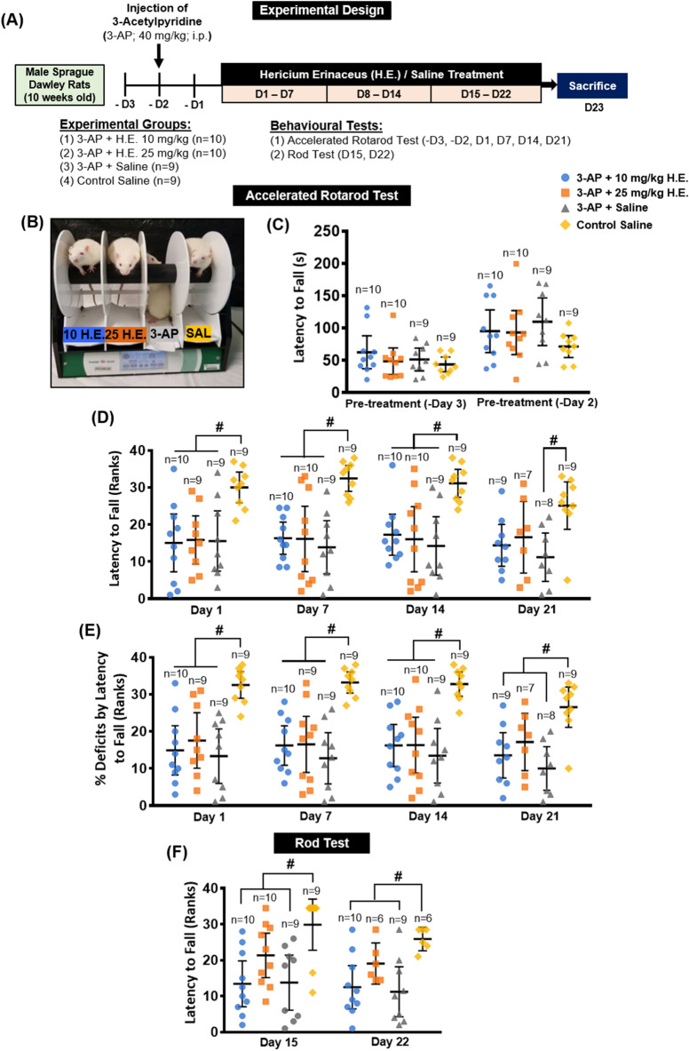
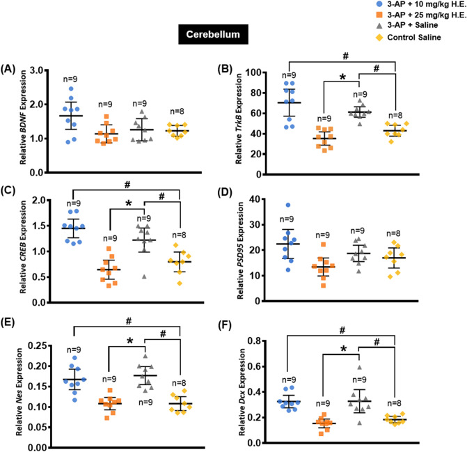
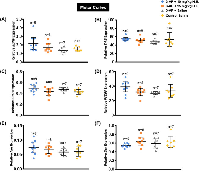
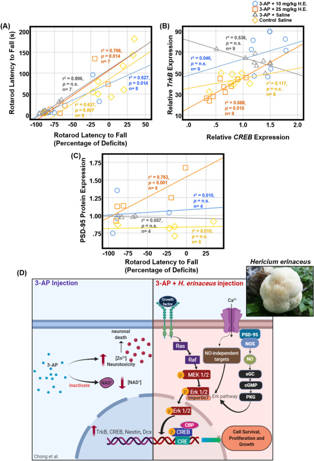
Cerebellar ataxia is a neurodegenerative disorder with no definitive treatment. Although several studies have demonstrated the neuroprotective effects of Hericium erinaceus (H.E.), its mechanisms in cerebellar ataxia remain largely unknown. Here, we investigated the neuroprotective effects of H.E. treatment in an animal model of 3-acetylpyridine (3-AP)-induced cerebellar ataxia. Animals administered 3-AP injection exhibited remarkable impairments in motor coordination and balance. There were no significant effects of 25 mg/kg H.E. on the 3-AP treatment group compared to the 3-AP saline group. Interestingly, there was also no significant difference in the 3-AP treatment group compared to the non-3-AP control, indicating a potential rescue of motor deficits. Our results revealed that 25 mg/kg H.E. normalised the neuroplasticity-related gene expression to the level of non-3-AP control. These findings were further supported by increased protein expressions of pERK1/2-pCREB-PSD95 as well as neuroprotective effects on cerebellar Purkinje cells in the 3-AP treatment group compared to the 3-AP saline group. In conclusion, our findings suggest that H.E. potentially rescued behavioural motor deficits through the neuroprotective mechanisms of ERK-CREB-PSD95 in an animal model of 3-AP-induced cerebellar ataxia.


Conflict of interest statement

The authors declare no competing interests.

Figures

Figure 1
Figure 1
Schematic representation of the experimental design for H. erinaceus treatments and a testing of cerebellar ataxia functions in rat model of 3-AP-induced cerebellar ataxia (A). Rats from each group were placed on the lane of the rotarod apparatus, respectively (B). Rats were trained on the rotarod and baseline performance was assessed on day -3 and day -2 prior to the 3-AP injection (C). Accelerated rotarod evaluation of the motor deficits by latency to fall (Rank) (D), percentage of deficit (Rank) (E), and rod test by latency to fall for the assessment of balance (F). The endpoints were rank transformed, and results were presented as mean individual data points with 95% confidence interval. All statistical values have been adjusted by Bonferroni correction for multiple comparisons. Indicators: # Significantly different from the non-3-AP control group. p values < 0.05.
Figure 2
Figure 2
Effects of 3-AP and H. erinaceus on the relative expression of neuroplasticity- and neurogenesis-related genes including BDNF (A), TrkB (B), CREB (C), PSD95 (D), Nes (E), Dcx (F) in the cerebellum of 3-AP + 10 mg/kg H.E., 3-AP + 25 mg/kg H.E., 3-AP + saline, and non-3-AP control groups. TrkB, CREB, Nes, and Dcx were significantly upregulated in 3-AP-induced ataxic rats without H. erinaceus treatment. Notably, 25 mg/kg H. erinaceus normalised the neuroplasticity-related gene expressions to the levels in the non-3-AP control. Relative expression was calculated by normalising the relative quantifications to the reference gene GAPDH as the ratio of the 2^CT(reference) and 2^CT(test). Indicators: # Significantly different from the non-3-AP control group. * Significantly different from the 3-AP + saline group. p values < 0.05.
Figure 3
Figure 3
Effects of 3-AP injection and H. erinaceus treatment on the expression of neuroplasticity- and neurogenesis-related genes including BDNF (A), TrkB (B), CREB (C), PSD95 (D), Nes (E), and Dcx (F) in the motor cortex of 3-AP + 10 mg/kg H.E., 3-AP + 25 mg/kg H.E., 3-AP + saline, and non-3-AP control groups. No significant changes were observed in the expression of neuroplasticity- and neurogenesis-related genes in the motor cortex among the different groups. Relative expression was calculated by normalising the relative quantifications to the reference gene GAPDH as the ratio of the 2^CT(reference) and 2^CT(test). Indicators: * Significantly different from the non-3-AP control group. # Significantly different from the 3-AP + saline group. p values < 0.05.
Figure 4
Figure 4
Western blot analysis of neuroplasticity-related proteins in cerebellar tissue from rats administered with 3-AP + 10 mg/kg H.E., 3-AP + 25 mg/kg H.E., 3-AP + saline and saline only (A). Note, dividing lines and white space representing the blots were cropped from different blots. Graphical representation of the effects of 3-AP and H. erinaceus administration on the expression of neuroplasticity-related proteins (A) including ERK1/2 (B), pERK1/2 (C), CREB (D), pCREB (E), Syp (F), and PSD95 (G). The expression of the target protein was normalised to the expression of GAPDH. Note, there were significant increases in the protein expressions of pERK1/2, pCREB and PSD95 in 3-AP + 25 mg/kg H.E. group compared to both the 3-AP + saline group and non-3-AP control group. Indicators: * Significantly different from the non-3-AP control group. # Significantly different from the 3-AP + saline group. p values < 0.05.
Figure 5
Figure 5
Effects of 3-AP and H. erinaceus administration on the morphological changes of Purkinje cells by haematoxylin and eosin staining. Notably, animals in the 3-AP + 10 mg/kg H.E. (A) and 3-AP + 25 mg/kg H.E. groups (B) showed Purkinje cells with regular and ordinary morphology that was similar to the non-3-AP control group. Interestingly, Purkinje cells in the 3-AP + saline group (C) were scattered with disturbed alignment and irregular appearance. The Purkinje cells in the non-3-AP control group (D) showed regular morphology and ordinary alignment between granular and molecular layers. The scale bars represent 50 μm. The Purkinje cell linear density of 3-AP + 10 mg/kg H.E. (1752 Purkinje cells per 58,729.942 μm total PC length, 30 sections from 6 animals), 3-AP + 25 mg/kg H.E. groups (1519 Purkinje cells per 50,799.906 μm total PC length, 25 sections from 5 animals), and the non-3-AP control group (1,295 Purkinje cells per 42,788.036 μm total PC length, 20 sections from 4 animals) showed significant differences compared to 3-AP + saline group (1,348 Purkinje cells per 51,171.87 μm total PC length, 25 sections from 5 animals) (E). Interestingly, there was a remarkable reduction of caspase-3 in 3-AP + 25 mg/kg H.E. animals compared to the 3-AP + saline group, indicating the neuroprotective effects of 25 mg/kg of H. erinaceus against apoptosis (F). A significant decrease of gene expression for PCP4 (G) and calbindin-D28k (H) was found in 3-AP + 25 mg/kg H.E. group compared to the 3-AP + saline and the non-3-AP control groups. Indicators: * Significantly different from the non-3-AP control group. # Significantly different from the 3-AP + saline group. p values < 0.05.
Figure 6
Figure 6
Scatter plots displaying the correlations between the variables related to the motor behavioural tests, as well as the cerebellar neuroplasticity- and neurogenesis-related relative gene and protein expressions. Notably, there were significant correlations between the accelerated rotarod behavioural data and the percentage of deficits in 3-AP + 10 mg/kg H.E., 3-AP + 25 mg/kg H.E., and non-3-AP control groups. No correlation was found between the accelerated rotarod behavioural data and the percentage of deficits in 3-AP + saline group (r2 = 0.896, p = n.s.), suggesting the behavioural motor coordination of accelerated rotarod was altered by 3-AP injection (A). In the 3-AP + 25 mg/kg H.E. group, the gene expression of TrkB was positively correlated with the CREB expression (B), and the PSD95 protein expression was positively correlated with the percentage of deficits by latency to fall (C). The hypothetical mechanism of neuronal cell death induced by 3-AP (left) and pathways of H. erinaceus on the role of pERK1/2-pCREB-PSD95 in neuroplasticity-related mechanisms (right). 3-AP reduces the intracellular concentration of NAD+ and interferes with Zn2+ homeostasis, which increases the Zn2+ neurotoxicity and lead to neuronal death. Upregulation of neuroplasticity and neurogenesis-related genes including TrkB, CREB, Nestin, and Dcx was stimulated by 3-AP to reverse neuronal death (left). After the injection of 3-AP followed by H. erinaceus, the tyrosine kinase receptors are hypothetically activated by NGF and followed by the recruitment of the cytoplasmic protein, Son of Sevenless (SOS). This triggers the activation of the Ras/guanosine triphosphate complex and initiates cytoplasmic kinase signal transduction cascades. MEK1/2 catalyses the phosphorylation of ERK1/2 at Tyr204/187 mediated by importin-7, followed by translocation from the cytoplasm to the nucleus,. ERK1/2 acts as an upstream regulator by catalysing the phosphorylation of various cytoplasmic and nuclear substrates that encode transcription factors and gene regulatory proteins, including CREB,. Additionally, the Ca2+ influx is through the NMDA receptors, which activates the neuronal nitric oxide synthase, that is mediated by PSD-95,. The induced nitric oxide activates ERK for the expression of neuroplasticity-related proteins, facilitated by cGMP and PKG (right). This hypothetical model was created by BioRender.com (D).

References

    1. Klockgether T, Paulson H. Milestones in ataxia. Mov Disord. 2011;26:1134–1141. doi: 10.1002/mds.23559. - DOI - PMC - PubMed
    1. Marsden JF. Chapter 17 - Cerebellar ataxia. In: Day BL, Lord SR, editors. Handbook of Clinical Neurology. Amsterdam: Elsevier; 2018. pp. 261–281. - PubMed
    1. Harding AE. Clinical features and classification of inherited ataxias. Adv Neurol. 1993;61:1–14. - PubMed
    1. Lim J, et al. A protein–protein interaction network for human inherited ataxias and disorders of Purkinje cell degeneration. Cell. 2006;125:801–814. doi: 10.1016/j.cell.2006.03.032. - DOI - PubMed
    1. Ferdinandusse S, et al. Ataxia with loss of Purkinje cells in a mouse model for Refsum disease. Proc. Natl. Acad. Sci. 2008;105:17712–17717. doi: 10.1073/pnas.0806066105. - DOI - PMC - PubMed

Publication types

MeSH terms