. 2020 Sep 10;11(1):4534.
doi: 10.1038/s41467-020-18306-x.

Resolution of R-loops by INO80 promotes DNA replication and maintains cancer cell proliferation and viability

Affiliations

Resolution of R-loops by INO80 promotes DNA replication and maintains cancer cell proliferation and viability

Lisa Prendergast et al. Nat Commun. .

Abstract

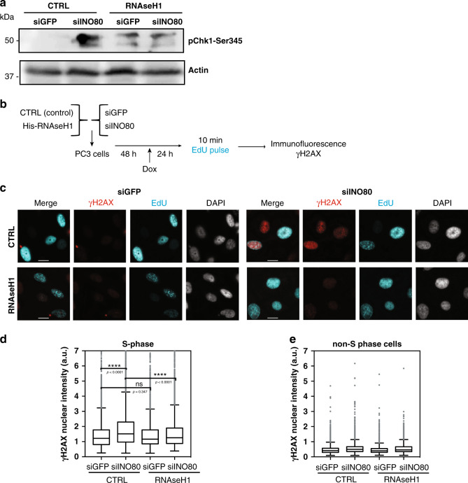
Collisions between the DNA replication machinery and co-transcriptional R-loops can impede DNA synthesis and are a major source of genomic instability in cancer cells. How cancer cells deal with R-loops to proliferate is poorly understood. Here we show that the ATP-dependent chromatin remodelling INO80 complex promotes resolution of R-loops to prevent replication-associated DNA damage in cancer cells. Depletion of INO80 in prostate cancer PC3 cells leads to increased R-loops. Overexpression of the RNA:DNA endonuclease RNAse H1 rescues the DNA synthesis defects and suppresses DNA damage caused by INO80 depletion. R-loops co-localize with and promote recruitment of INO80 to chromatin. Artificial tethering of INO80 to a LacO locus enabled turnover of R-loops in cis. Finally, counteracting R-loops by INO80 promotes proliferation and averts DNA damage-induced death in cancer cells. Our work suggests that INO80-dependent resolution of R-loops promotes DNA replication in the presence of transcription, thus enabling unlimited proliferation in cancers.


Conflict of interest statement

The authors declare no competing interests.

Figures

Fig. 1
Fig. 1. R-loops slow replication rate in INO80-depleted cells.
a Schematic representation of the experimental approach. PC3 cells were transfected with esiRNAs against either GFP or INO80. Three days later, cells were treated with α-amanitin (α-a) or cordycepin (crd) for 3 h or left untreated as control and then subjected to fibre labelling analysis. b Representative images of spread fibres from each condition. Similar results were obtained in five independent experiments (cordycepin treatment—2). c Distribution of fork speed rates in INO80-proficient (siGFP) and INO80-deficient (siINO80). Data is from five independent experiments (in cordycepin-treated cells—2), at least 250 fibres were measured per condition in each experiment. ****p-value < 0.0001, (two- tailed unpaired Student’s t-test). d Schematic representation of the experimental setup used. PC3 cells were co-transfected with esiRNAs against either GFP (siGFP) or INO80 (siINO80) along with either a control (CTRL) or RNase H1-overexpressing (RNAseH1) vector. Two days later RNAse H1 expression was induced by doxycycline for 24 h. Cells were labelled with CldU for 5 min followed by IdU pulse for 20 min and subjected to DNA fibre labelling analysis. e Representative images of spread fibres from each condition. Similar results were obtained in four independent experiments. f Distribution of fork speed rates (kilobase/min) in siGFP and INO80-deficient cells transfected with control or RNAse H1 overexpression plasmids. Data are from 4 independent experiments, at least 250 fibres were measured per condition in each experiment. ****p-value < 0.0001, (two-tailed unpaired Student’s t-test). g Schematic representation of the experimental setup. Cells were co-transfected and induced as in d and prior to labelling were treated with 5 µM vorinostat for 6 h. h Representative images of spread fibres from each condition. Similar results were obtained in three independent experiments. i Distribution of fork rates from (h, at least 250 fibres were measured per condition in each experiment; ns non-significant, ****p-value < 0.0001, *p-value < 0.05, (two-tailed unpaired Student’s t-test). c, f, i Kruskal–Wallis test p-value was < 0.0001. Data is presented as Tukey boxplot (box representing first quartile, median and third quartile, whiskers 1.5 times interquartile range).
Fig. 2
Fig. 2. Fork asymmetry in Ino80-depleted cells depends on R-loops.
a Schematic representation of the experimental setup used. PC3 cells were co-transfected with esiRNAs against either GFP (siGFP) or INO80 (siINO80) along with either a control (CTRL) or RNAse H1-overexpressing (RNAse H1) vector. Two days later RNAse H1 expression was induced by doxycycline for 24 h. Cells were labelled with CldU for 20 min (red) followed by IdU pulse for 20 min and subjected to DNA fibre labelling analysis. b Representative pairs of sister replication forks were assembled from different fields of view and were arbitrarily centred on the position of origin. Scale bar 5 μm. Similar distribution of paired forks was observed in three independent experiments. c Scatter plots of the distances covered by right-moving and left-moving sister forks during the CldU pulse in Ino80-proficient or deficient cells expressing or not RNAse H1. The central areas delimited with grey lines contain sister forks with less than a 25% length difference. The percentage of symmetric forks is indicated. d Relative fork asymmetry. Fork asymmetry is expressed as the ratio of the longer arm to the shorter one for each pair of sister replication forks, ****p-value < 0.0001; n.s non-significant (two-tailed unpaired Student’s t- test). Numbers above boxes indicate the median of the ratio of the longer to shorter arm. Data is presented as Tukey boxplot.
Fig. 3
Fig. 3. Replication stress-induced DNA damage in INO80-deficient cells is caused by R-loops.
a PC3 cells were transfected with control vector (CTRL) and either esiRNA against GFP (lane 1) or INO80 (lane 2) or transfected with RNAse H1-expressing plasmid (RNAseH1) and esiRNA against GFP (lane 3) or INO80 (lane 4). Forty-eight hours later cells were induced with doxycycline and 24 h later analyzed by Western with an antibody against pChk1-Ser345. Similar results were obtained in two independent experiments. b Schematic representation of the experimental setup used. PC3 cells were co-transfected with esiRNAs against either GFP (siGFP) or INO80 (siINO80) along with either a control (CTRL) or RNAse H1-overexpressing (RNAseH1) vector. Two days later RNase H1 overexpression was induced by doxycycline for 24 h. To distinguish cells in S-phase, cells were labelled with 25 µM EdU, fixed and stained with an antibody against γH2AX and “clicked” with Alexa Fluor 488 azide. c Representative images of cells as in b. Scale bar 10 µm d Distribution of nuclear γH2AX staining intensities in S-phase cells; ****p-value < 0.0001, ns nonsignificant (two-tailed unpaired Student’s t-test). At least 500 cells were measured per condition in each of three independent experiments. Data is presented as Tukey boxplot. e Distribution of nuclear γH2AX staining intensities in non-S-phase cells. Data in d, e are from three independent experiments following normalization (to median intensity of entire population of siGFP/CTRL sample in each experiment). Tukey boxplot is used. Source data are provided as a Source data file.
Fig. 4
Fig. 4. R-loops accumulate in INO80-depleted cells.
a Schematic representation of the experimental setup used. PC3 cells were co-transfected with esiRNAs against GFP (siNT) or INO80 (siINO80). Cells were pulsed with EdU for 15′ and immunofluorescence was carried out using the S9.6 antibody against R-loops. b Representative immunoblot of total extracts from PC3 cells transfected with esiRNAs against GFP (siGFP) or INO80 (siINO80) with an antibody against INO80 3 days after transfection. Similar results were obtained in five independent experiments. c Representative confocal deconvolved images EdU positive and EdU negative cells immunostained with S9.6 n = 3 biological replicates were quantified. Scale bar is 10 μM. d Total nuclear fluorescence intensity of R-loops in 3D volume, in control and INO80-depleted cells. DAPI was used as a mask to measure total fluorescence intensity of R-loops in 3D nuclear volume. Number of cells analyzed in each condition: siNT: n = 133 cells; siINO80: n = 151 cells. Data are from three independent experiments. p-value = 0.0002, (two-tailed unpaired Student’s t-test). e Total nuclear fluorescence intensity of R-loops in 3D volume, in control (siNT) and INO80-depleted (siINO80) cells outside (non-EdU) and inside (EdU) S-phase. DAPI was used as in d. Number of cells analyzed in each condition: siNT(non-EdU): n = 91; siINO80(non-EdU): n = 117; siNT(EdU): n = 42; siINO80(EdU): n = 26. Data are from three independent experiments. ***p-values = 0.0003 *p-value=0.0117, (two-tailed unpaired Student’s t-test). f Representative immunoblot of PC3 cells transduced with control shRNA (ShScramble) RNA and four different shRNAs targeting INO80 (shINO80-1-4) following experimental setup shown in Supplementary Fig. 4b. Similar results were obtained in 4 independent experiments. g DRIP-qPCR was performed on the indicated shRNA-treated samples using the S9.6 antibody. Values for each region tested were normalized over shControl after correction for input DNA levels. Mock IP was conducted in shControl and shINO80 samples in the absence of S9.6 antibody. Data from three independent biological replicates are presented as mean values ± SD. Source data are provided as a Source data file.
Fig. 5
Fig. 5. Colocalization of INO80 with R-loops by STED nanoscopy.
a Single image plane of confocal and STED imaging of PC3 cells. Cells were pulsed with EdU for 15′ and immunostained with S9.6 and anti-INO80 antibodies and imaged using confocal (EdU) and STED (INO80 and S9.6). STED imaging pixel size is 11 nm in XY. Panel 1, 2 and 3 are magnified regions where INO80 and S9.6 co-localize. Scale bar =  100 nm n = 6 independent cells were analysed from three replicates b 3D volume surface model of INO80 and S9.6. Transparency of the INO80 volume (green) has been increased to enable visualization of overlapping regions (yellow). Similar results were obtained from six independent cells. c Upper panel. The number of R-loops per cell was determined in six independent EdU positive cells. Thresholds were generated based on fluorescence intensity within the Hoechst stained nuclear volume and excluded objects that were below 10 voxels in size as background. The number of R-loops (S9.6 volumes) which shared co-localizing voxel volumes with INO80 were quantified. Lower panel. Bar-graph indicating the percent of R-loops colocalizing with INO80 relative to the total amount of R-loops in each cell. df Comparative analysis of R-loop intensity sum d, voxel volume e and length f in colocalizing (coloc) or non-colocalizing (non-coloc) with INO80 in Cell 1 n = 250 non co-localising R-loops, n = 168 colocalising R-loops. (****adjusted p-value < 0.0001; one way ANOVA). Analysis of the cells 2–6 is in Supplementary Fig. 9. Data are presented as mean values ± Standard deviation (SD)”.
Fig. 6
Fig. 6. R-loops associate with INO80 genome-wide and promote INO80 binding to chromatin.
a Smoothed scatterplot showing the pairwise correlation between DRIP-seq (R-loops) and INO80 ChIP-seq abundances at gene bodies in mESCs. b Genomic enrichment profile of INO80 and R-loops (DRIP) signals across the beta-actin (Actb) gene in mouse ESCs. c Heat map for model parameters of ChromHMM, indicating the relative emission probability of each mark/feature to each state. d Genome-wide enrichment of DRIP-seq (R-loops) and INO80 ChIP-seq peaks at 200 bp binning resolution across different chromatin states based on marks shown in Fig. 4c. Columns indicate the relative percentage of the genome represented by each chromatin state and relative fold enrichment for different types of annotation. INO80 + DRIP: overlapping INO80 and DRIP-seq peaks; DRIP: DRIPseq peaks not overlapping with INO80 peaks; INO80: INO80 peaks not overlapping with DRIPseq peaks; NoDRIP+NoINO80: regions of the genome lacking both DRIPseq and INO80 peaks; TES Transcription End Site; TSS Transcription Start Site. e Schematic representation of the differential salt fractionation setup used. PC3 cells stably expressing either control vector or a Tet-inducible RNase H1-expressing vector were treated by doxycycline for 24 h. Sequential subcellular fractionated lysates were isolated in low salt (150 mM), high salt (600 mM) and from benzonase-digested pellet to release the tightly bound chromatin-associated factors. Fractions analyzed by immunoblot for INO80 and histone H3. f Immunoblot of subcellular fractions from control and RNHaseH1 expressing cells from a representative chromatin fractionation experiment. Histone H3 was used as a marker for chromatin enrichment. INO80 and H3 were analyzed from the same gel. INO80* indicates lighter exposure of the INO80 immunoblot. N = 3 biological replicates were performed with similar results. g Bar graph indicating the change in INO80 enrichment in the chromatin fraction between control and RNAse H1 overexpressing cells. The amount of INO80 detected in the chromatin fraction of the control cells was set arbitrarily to 100%. Data are from three independent experiments. **p-value = 0.0027 two-tailed unpaired Student’s t-test). Data are presented as mean values ± SD. Quantification by ImageJ. Source data are provided as a Source data file.
Fig. 7
Fig. 7. Dynamic R-loop turnover at the LacO locus by INO80 tethering.
a Schematic representation of the experimental assay. LacI-eGFP tagged INO80E or RNHseH1 were transfected into U2OS cells harbouring a 256xLacO array. Immunostaining with S9.6 after 24 h allows visualization of R-Loops at the LacO locus. b Representative immunostained images of LacO cells transfected with eGFP-LacI, eGFP-LacI- INO80E, eGFP-LacI-RNHaseH1. Scale bar is 10 µm. N = 4 biological replicates were performed with similar results c Boxplot showing the S9.6 signal intensity relative to the underlying eGFP-LacI-tagged proteins intensity at the LacO locus in the respective conditions, as described in a. Plot shown is min to max values with line at median. Number of cells per condition shown: eGFP = 97; RNase H1 = 116; INO80E = 100. N = 3 experiments ****adjusted p-value < 0.0001; one way ANOVA test. d Schematic representation of the experimental assay. LacO-carrying U2OS cells were co-transfected with RBD-DsRed and either LacI-eGFP or LacI-eGFP-INO80E plasmids and live cell imaging carried out in Z stacks imaged every 6 min. e Representative live images of LacO cells transfected with LacI-eGFP or LacI-eGFP-INO80E n = 3 biological replicates gave similar results. Scale bar is 10 µm. f Images of selected time-points are shown from a representative live imaging experiment. Merge shows LacO co-localisation with LacI-eGFP or LacI-INO80E cell during 48 min of the time course analysis for RBD-DsRed and LacI-tagged proteins. Merged images of whole cells are in Supplementary Figure 11d. Montage is a zoom of the LacO site from the representative cell in e. g Upper panel: formula for calculating Fold-Change in Intensity (FC-I). Intensity change of the RBD-DsRed signal normalized over the underlying eGFP signal at the locus was measured at 6-minute intervals throughout the course of the experiment for a total of 1500 min. FC-I was calculated as the normalized RBD-DsRed signal at each timepoint relative to the previous timepoint. Lower panel: Total, negative and positive FC-I(log2) values for RBD-DsRed signal in LacI-eGFP and LacI-INO80E cells. The total number of FC-I(log2) values was approximately 900 for both LacI-eGFP and LacI-INO80E. Positive FC-I(log2) values are 70–75% of the total. Negative FC-I(log2) are 25-30% of the total. N = 5 independent live cells quantified for each condition. ****adjusted p-value < 0.0001; ANOVA with Kruskal–Wallis test.
Fig. 8
Fig. 8. RNAse H1 overexpression rescues cancer cell proliferation in the absence of INO80.
ad Cells from the indicated lines were co-transfected with control (siGFP) or INO80 targeting (siINO80) esiRNAs along with either a control (CTRL) or RNAse H1-overexpressing (oeRNAse H1) vector. Proliferation was monitored by cell counting 96 h after transfection. Expected values to test for independent effect of INO80 depletion and overexpression of RNAse H1 in cell growth are calculated according to, as: V = [%growth siINO80] × [%growth oeRNAse H1]. Data are from 3 independent experiments. Exact p values: Fig. 8a = 0.1 for siRNaseH1 and 0.99 for siINO80 vs siGFP, Fig. 8bp-value = siGFP vs siINO80 = 0.047, p-value siGFP vs siINO80 = 0.007; p-value siINO80-siRNaseH1 vs predicted = 0.05. Figure 8c: p-value siGFP vs siINO80 = 0.009, siINO80-siRNaseH1 vs predicted = 0.046 Fig. 8d: p-value siGFP vs siRNaseH1 = 0.0005, p-value siGFP vs siINO80 > 0.0001, siINO80-siRNaseH1 vs predicted 0.0245 *p value < 0.05; **p-value < 0.01, ***p-value < 0.005; two-tailed unpaired Student’s t-test. Data are presented as mean values ± SEM e Cell death analysis. PC3 cells transfected with control (siGFP), RNase H1-targeting (siRNaseH1) or INO80-targeting (siINO80) siRNAs for 24 h were treated with increasing concentrations of DL-Dopa for further 7 days incubation and analysed for cell growth (Supplementary Fig. 9f) and cytotoxicity. Cell death was calculated for cytotoxicity fluorescence values normalized to the respective relative cell growth. Fold change cell death values were calculated by setting untreated control cells arbitrarily to 1. Concentrations used for DL-Dopa inhibitor: non-treated (−), 1 μM, 2 μM and 5 μM. Data are presented as mean values ± SD, measure of centre is mean. Data are from three independent experiments. P values calculated by unpaired two-tailed t-test. (siGFP v siINO80 P value **p = 0.0036, siGFP 2 μM v siINO80 2 μM P value *p = 0.0464, siGFP 5 μM v siRNAseH1 5 μM P value *p = 0.0156.

References

    1. Garcia-Muse T, Aguilera A. R Loops: from physiological to pathological roles. Cell. 2019;179:604–618. - PubMed
    1. Sollier J, Cimprich KA. Breaking bad: R-loops and genome integrity. Trends Cell Biol. 2015;25:514–522. - PMC - PubMed
    1. Gaillard H, Aguilera A. Transcription as a threat to genome integrity. Annu Rev. Biochem. 2016;85:291–317. - PubMed
    1. Tuduri S, et al. Topoisomerase I suppresses genomic instability by preventing interference between replication and transcription. Nat. Cell Biol. 2009;11:1315–1324. - PMC - PubMed
    1. Hamperl S, Bocek MJ, Saldivar JC, Swigut T, Cimprich KA. Transcription-replication conflict orientation modulates R-Loop levels and activates distinct DNA damage responses. Cell. 2017;170:e719. - PMC - PubMed

Publication types

Substances