

Metastatic melanoma presenting as a breast mass - role of radiologist as a clinician
- PMID: 32913479
- PMCID: PMC7473873
- DOI: 10.1016/j.radcr.2020.08.009
Metastatic melanoma presenting as a breast mass - role of radiologist as a clinician
Abstract
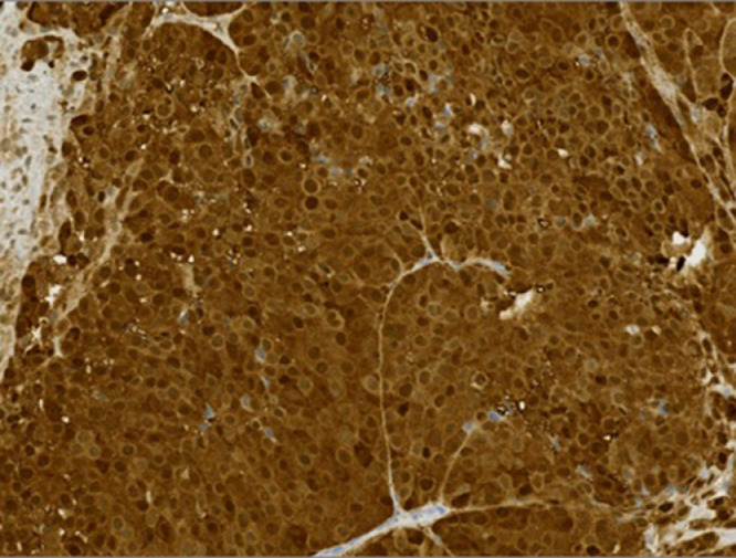
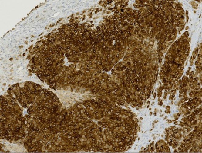
Breast tissue can be the host of not only many benign and malignant tumors but can also be a metastatic site for various tumors such as leukemia, lung cancer, and melanoma. This report describes an unusual case of a 43-year-old female who presented with a new palpable breast lump and several similar extramammary lumps on her skin. A melanoma panel, consisting of S100, HMB45, and Melan-A stains, was included in the pathology evaluation due to diagnostic suspicion of the radiologist and revealed metastatic melanoma. This case highlights the importance of detailed history and relevant physical exam as well as clinical and imaging correlation. It serves as a reminder to radiologists to include metastatic melanoma in the differential of suspicious subcutaneous breast masses, especially in patients with multiple subcutaneous lumps in the body or abnormal skin findings.
© 2020 The Authors.
Figures
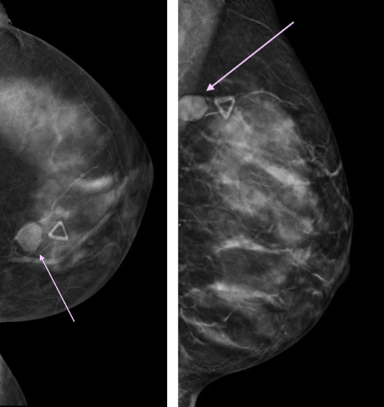
 pink arrow), corresponding to the area of the palpable abnormality (
 skin marker).
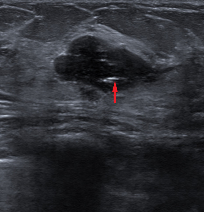
 red arrow) consistent with biopsy proven fibroadenoma. (Color version of figure is available online.)
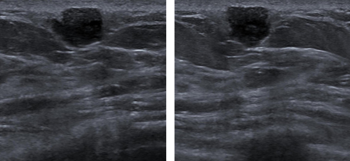
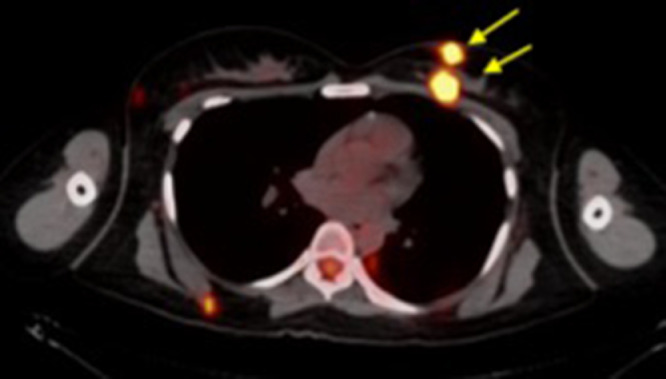
 yellow arrows), previously identified on ultrasound at 10-o'clock were also FDG avid. (Color version of figure is available online.)
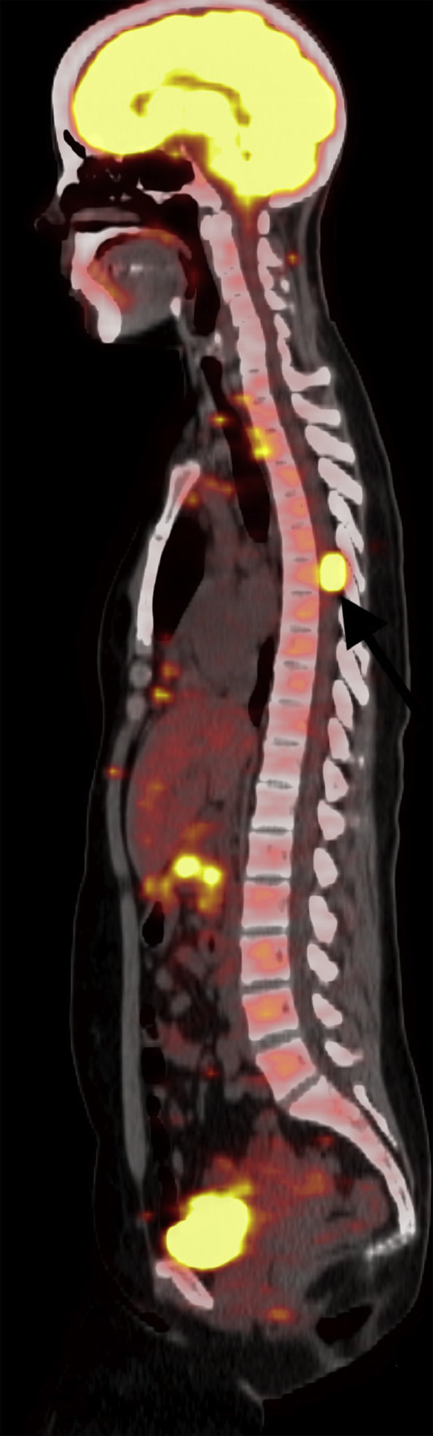
 black arrow).
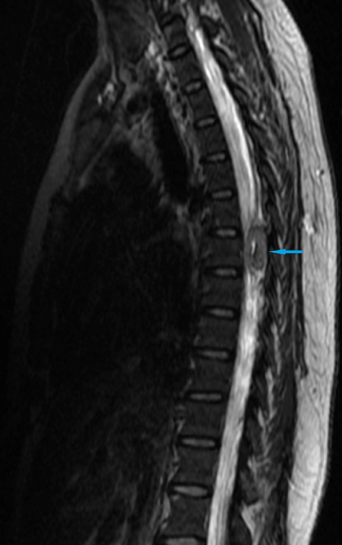
 blue arrow). (Color version of figure is available online.)
References
-
- Kurul S, Tas F, Buyukbabani N, Mudun A, Baykal C, Camlica H. Different manifestations of malignant melanoma in the breast: a report of 12 cases and a review of the literature. Jpn J Clin Oncol. 2005;35(4):202–206. - PubMed
-
- Bassi F, Gatti G, Mauri E, Ballardini B, De Pas T, Luini A. Breast metastases from cutaneous malignant melanoma. Breast. 2004;13(6):533–535. - PubMed
-
- Ravdel L, Robinson WA, Lewis K, Gonzalez R. Metastatic melanoma in the breast: a report of 27 cases. J Surg Oncol. 2006;94(2):101–104. - PubMed
Publication types
LinkOut - more resources
Full Text Sources
