The etiological role of endoplasmic reticulum stress in acute lung injury-related right ventricular dysfunction in a rat model
- PMID: 32913512
- PMCID: PMC7476135
The etiological role of endoplasmic reticulum stress in acute lung injury-related right ventricular dysfunction in a rat model
Abstract
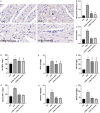
This study aimed to ascertain whether endoplasmic reticulum (ER) stress participates in acute lung injury (ALI) and related right ventricular dysfunction (RVD) as well as to explore the underlying mechanisms of these conditions. A single intratracheal instillation of lipopolysaccharide (LPS) (10 mg/kg) was used to establish the RVD model. The ER stress inhibitor, 4-PBA (500 mg/kg), was administered using a gavage 2 hours before and after the LPS treatment for prevention and treatment, respectively. At 12 hours post-LPS exposure, mRNA and protein expressions of ER stress-specific biomarkers, glucose regulating protein 78 (GRP78) and CCAAT/enhancer binding protein homology (CHOP), were significantly upregulated. This effect was inhibited by both 4-PBA prevention and treatment. In addition, echocardiography showed that 4-PBA improved the LPS-induced abnormality in the tricuspid annular plane systolic excursion (TAPSE) and the right ventricular end-diastolic diameter (RVEDD), however not in the pulmonary artery acceleration time (PAAT). Furthermore, hematoxylin and eosin staining (HE) and terminal transferase dUTP nick end labeling (TUNEL) assays revealed that the proportion of proapoptotic cells was higher in RVD rats. This was prominently ameliorated by 4-PBA treatment. Moreover, 4-PBA had a similar reverse effect on the LPS-induced increase in the Bax/Bcl-2 ratio, caspase-12, and caspase-3 expressions as revealed by western blotting. Furthermore, 4-PBA improved LPS-induced right ventricle (RV) myeloperoxidase (MPO)-positive neutrophil infiltration percentage, inhibited nuclear factor kappa B (NF-κB) activity, and reduced the expressions of inflammatory cytokines, TNF-α, IL-1β, and IL-6, in serum and RV. Taken together, our results indicated that ER stress-mediated apoptosis and inflammation might contribute to the development of ALI-related RVD induced by intratracheal LPS instillation. Gavage-administered 4-PBA could improve right ventricle (RV) systolic dysfunction and dilation, plausibly by blocking ER stress.
Keywords: 4-phenyl butyric acid; Endoplasmic reticulum stress; acute lung injury; apoptosis; inflammation; right ventricular dysfunction.
AJTR Copyright © 2020.
Conflict of interest statement
None.
Figures


Similar articles
-
[Protective effect of sodium 4-phenylbutyrate on rats with acute respiratory distress syndrome related right ventricular dysfunction by alleviating endoplasmic reticulum stress].Zhonghua Wei Zhong Bing Ji Jiu Yi Xue. 2019 Oct;31(10):1269-1274. doi: 10.3760/cma.j.issn.2095-4352.2019.10.017. Zhonghua Wei Zhong Bing Ji Jiu Yi Xue. 2019. PMID: 31771727 Chinese.
-
Roles of apoptosis and inflammation in a rat model of acute lung injury induced right ventricular dysfunction.Biomed Pharmacother. 2018 Dec;108:1105-1114. doi: 10.1016/j.biopha.2018.09.115. Epub 2018 Sep 29. Biomed Pharmacother. 2018. PMID: 30372811
-
4-PBA inhibits LPS-induced inflammation through regulating ER stress and autophagy in acute lung injury models.Toxicol Lett. 2017 Apr 5;271:26-37. doi: 10.1016/j.toxlet.2017.02.023. Epub 2017 Feb 27. Toxicol Lett. 2017. PMID: 28245985
-
[Role of inflammation and apoptosis in right ventricular dysfunction induced by injurious mechanical ventilation in rats].Zhonghua Wei Zhong Bing Ji Jiu Yi Xue. 2022 May;34(5):519-524. doi: 10.3760/cma.j.cn121430-20210401-00482. Zhonghua Wei Zhong Bing Ji Jiu Yi Xue. 2022. PMID: 35728855 Chinese.
-
[Effects of acute respiratory distress syndrome induced by endotoxin on the right ventricular function in rats].Zhonghua Wei Zhong Bing Ji Jiu Yi Xue. 2018 Mar;30(3):204-208. doi: 10.3760/cma.j.issn.2095-4352.2018.03.003. Zhonghua Wei Zhong Bing Ji Jiu Yi Xue. 2018. PMID: 29519276 Chinese.
References
-
- Ma S, Wang X, Yao J, Cao Q, Zuo XR. Roles of apoptosis and inflammation in a rat model of acute lung injury induced right ventricular dysfunction. Biomed Pharmacother. 2018;108:1105–1114. - PubMed
LinkOut - more resources
Full Text Sources
Research Materials
Miscellaneous
