. 2020 Aug 15;12(8):4467-4477.
eCollection 2020.

Elabela alleviates myocardial ischemia reperfusion-induced apoptosis, fibrosis and mitochondrial dysfunction through PI3K/AKT signaling

Affiliations

Elabela alleviates myocardial ischemia reperfusion-induced apoptosis, fibrosis and mitochondrial dysfunction through PI3K/AKT signaling

Peng Yu et al. Am J Transl Res. .

Abstract

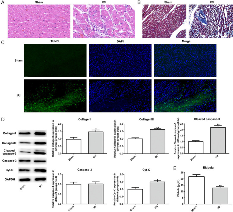
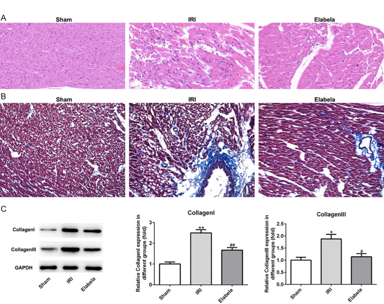
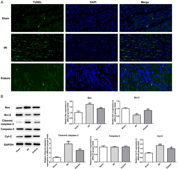
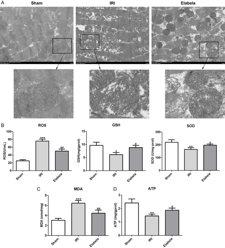
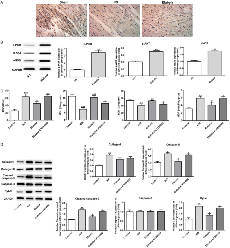
Myocardial ischemia/reperfusion (I/R) injury is a common cardiovascular disease with high morbidity and mortality globally, which derives from acute myocardial infarction and coronary artery disease. Elabela has been proved to bind to apelin receptors in the heart. The present study aimed to investigate the protective effects of Elabela in myocardial I/R injury and illustrating the potential mechanisms. In this study, the rat I/R model was established in vivo. Following treatment with Elabela, the histopathological changes of heart tissue were evaluated by the hematoxylin and eosin- or Masson's trichrome staining. Apoptosis of heart tissue was examined using TUNEL staining. The expression of type I or III collagen and apoptosis-associated proteins was measured using western blotting. Moreover, myocardial ultrastructure in myocardium was detected via electron microscopy analysis. H9c2 cells were treated with hypoxia/reoxygenation (H/R) to mimic the myocardial I/R injury in vitro. After treatment with Elabela or Elabela combined with LY294002, the levels of oxidative stress and apoptosis were examined. The results revealed that Elabela significantly improved the pathological changes of rat myocardial tissues induced by I/R. Additionally, Elabela treatment reduced cardiomyocyte I/R induced fibrosis and apoptosis as well as ameliorated mitochondrial dysfunction in animal and cells. Within inhibition of PI3K pathway, the protective effects of Elabela was reversed. Taken together, these findings demonstrated that Elabela could protect against fibrosis, apoptosis and oxidative stress via PI3K/ATK signaling pathway in cardiac ischemia reperfusion.

Keywords: Cardiac ischemia reperfusion; Elabela; apoptosis; fibrosis; mitochondrial function.


Conflict of interest statement

None.

Figures

Figure 1
Figure 1
The level of Elabela was reduced after myocardial ischemia/reperfusion injury. A. Heart H&E staining from sham and ischemia reperfusion injury heart tissue. Scale bar: 20 μm. B. Myocardium underwent Masson’s trichrome staining. Red parts indexed cardiomyocytes and blue parts indexed fibrosis. Scale bar: 20 μm. C. TUNEL-positive (green) cardiomyocytes were evaluated by double staining with TUNEL (green) and 4’, 6’-diamidino-2-phenylindole (blue). The number of TUNEL-positive cardiomyocytes at infarct area after reperfusion was significantly increased compared to the sham group. Scale bar: 50 μm. D. Expression levels of Bcl-2, Bax, cleaved-caspase3, caspase-3 and Cyt-c tested by western blotting in vivo. E. Levels of Elabela in blood of rats. All results were obtained from at least three independent experiments. All numerical data were presented as the mean ± standard deviation. IRI, ischemia reperfusion injury. *P < 0.05, **P < 0.01 versus sham group.
Figure 2
Figure 2
Elabela decreased cardiac fibrosis after myocardial ischemia/reperfusion injury. A. Heart H&E staining from sham and ischemia reperfusion injury heart tissue. Scale bar: 20 μm. B. Myocardium underwent Masson’s trichrome staining. Red parts indexed cardiomyocytes and blue parts indexed fibrosis. Scale bar: 20 μm. C. Expression levels of type I and type III collagen. IRI, ischemia reperfusion injury. All results were obtained from at least three independent experiments. All numerical data were presented as the mean ± standard deviation. *P < 0.05, **P < 0.01 versus sham group; #P < 0.05, ##P < 0.01 versus IRI group.
Figure 3
Figure 3
Elabela alleviated apoptosis triggered by cardiac ischemia/reperfusion. A. TUNEL-positive (green) cardiomyocytes were evaluated by double staining with TUNEL (green) and 4’, 6’-diamidino-2-phenylindole (blue). Scale bar: 20 μm. B. Expression levels of Bcl-2, Bax, cleaved-caspase3, caspase-3 and Cyt-c were measured using western blotting. IRI, ischemia reperfusion injury. All results were obtained from at least three independent experiments. All numerical data were presented as the mean ± standard deviation. *P < 0.05, **P < 0.01, ***P < 0.001 versus sham group; #P < 0.05 versus IRI group.
Figure 4
Figure 4
Elabela ameliorated mitochondrial dysfunction. (A) Transmission electron microscopic images from rat cardiac tissues. Scale bar: 2.0 μm. (B) Regular biomarkers including reactive oxygen species (ROS), glutathione (GSH), superoxide dismutase (SOD) and (C) malondialdehyde (MDA) were determined by commercial kit for oxidative stress. (D) Production of ATP was subjected by commercial kit. All results were obtained from at least three independent experiments. All numerical data were presented as the mean ± standard deviation. *P < 0.05, **P < 0.01, ***P < 0.001 versus sham group; #P < 0.05, ##P < 0.01 versus IRI group.
Figure 5
Figure 5
Elabela modulated the apoptosis and fibrosis by activating PI3K/AKT signaling pathway. A. The expression of phosphorylation-AKT was detected using immunohistochemical staining. B. Expression levels of eNOS, phosphorylation PI3K and AKT were examined using western blot analysis in vivo. **P < 0.01 versus IRI group. H9c2 cells were subjected to high glucose and hypoxia/reperfusion treatment. C. Levels of ROS, GSH, SOD and MDA were detected by commercial kits. D. Levels of type I and type III collagen, cleaved-casepase3, caspase3 and Cyt-c were assessed using western blot analysis in high glucose and hypoxia/reperfusion (H/R)-treated H9c2 cells. All results were obtained from at least three independent experiments. All numerical data were presented as the mean ± standard deviation. *P < 0.05, **P < 0.01, ***P < 0.001 versus control group; #P < 0.05, ##P < 0.01, ###P < 0.001 versus H/R group; ΔP < 0.05 versus Elabela group.

References

    1. Zhou H, Ma Q, Zhu P, Ren J, Reiter RJ, Chen Y. Protective role of melatonin in cardiac ischemia-reperfusion injury: from pathogenesis to targeted therapy. J Pineal Res. 2018;64 - PubMed
    1. Anselmi A, Abbate A, Girola F, Nasso G, Biondi-Zoccai GG, Possati G, Gaudino M. Myocardial ischemia, stunning, inflammation, and apoptosis during cardiac surgery: a review of evidence. Eur J Cardiothorac Surg. 2004;25:304–311. - PubMed
    1. Hausenloy DJ, Yellon DM. Myocardial ischemia-reperfusion injury: a neglected therapeutic target. J Clin Invest. 2013;123:92–100. - PMC - PubMed
    1. Matute-Bello G, Martin TR. Science review: apoptosis in acute lung injury. Crit Care. 2003;7:355–358. - PMC - PubMed
    1. Chng SC, Ho L, Tian J, Reversade B. ELABELA: a hormone essential for heart development signals via the apelin receptor. Dev Cell. 2013;27:672–680. - PubMed

LinkOut - more resources