Symptomatic vertebral hemangioma during pregnancy period: A case series and systematic literature review
- PMID: 32913613
- PMCID: PMC7459382
- DOI: 10.4081/or.2020.8685
Symptomatic vertebral hemangioma during pregnancy period: A case series and systematic literature review
Abstract
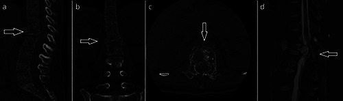
Vertebral Hemangioma (VH) is a benign tumor usually symptomless and discovered incidentally. Pregnancy, because of several hormonal and physiologic changes, is a recognized risk factor coinciding with the development of a rapid onset of neurological symptoms in patients affected by VH. In the Literature, sporadic cases of neurological symptoms have been described, which occurred during pregnancy, but only rarely the onset of symptoms was reported after pregnancy and childbirth. Usually surgical treatment is reserved for severe cases with rapid onset of neurological symptoms. However, the use of conservative treatments is still a topic of debate In the present study, we report a series of patients affected by VH become symptomatic during or after pregnancy along with a systematic review of the Literature.
Keywords: paraparesis; pregnancy; spine; spine tumors; vertebral hemangioma.
©Copyright: the Author(s).
Conflict of interest statement
Conflict of interest: the authors declare no potential conflict of interest.
Figures


Similar articles
-
Pregnancy related symptomatic vertebral hemangioma.Ann Indian Acad Neurol. 2014 Jan;17(1):120-2. doi: 10.4103/0972-2327.128577. Ann Indian Acad Neurol. 2014. PMID: 24753678 Free PMC article.
-
Acute spinal cord compression caused by vertebral hemangioma.Spine J. 2004 Sep-Oct;4(5):595-600. doi: 10.1016/j.spinee.2003.08.034. Spine J. 2004. PMID: 15363434 Review.
-
Surgical management of symptomatic vertebral hemangiomas: A case report and literature review.Surg Neurol Int. 2021 Feb 17;12:56. doi: 10.25259/SNI_752_2020. eCollection 2021. Surg Neurol Int. 2021. PMID: 33654559 Free PMC article. Review.
-
[Vertebral hemangioma symptomatic during pregnancy. A case report and review of the literature].J Gynecol Obstet Biol Reprod (Paris). 1997;26(1):90-4. J Gynecol Obstet Biol Reprod (Paris). 1997. PMID: 9091551 Review. French.
-
Pregnancy-related vertebral hemangioma. Case report, review of the literature, and management algorithm.Neurosurg Focus. 2005 Sep 15;19(3):E7. doi: 10.3171/foc.2005.19.3.8. Neurosurg Focus. 2005. PMID: 16190606 Review.
Cited by
-
Recurrent symptomatic vertebral hemangioma in pregnancy managed with decompression and vertebroplasty.Surg Neurol Int. 2021 Apr 8;12:150. doi: 10.25259/SNI_34_2021. eCollection 2021. Surg Neurol Int. 2021. PMID: 33948320 Free PMC article.
-
Lower-limb progressive paraparesis management and diagnosis overview in a pregnant woman with vertebral haemangioma.Womens Health (Lond). 2022 Jan-Dec;18:17455057221099018. doi: 10.1177/17455057221099018. Womens Health (Lond). 2022. PMID: 35574823 Free PMC article.
References
-
- Demirkale İ, De Iure F, Terzi S, Gasbarrini A. Aggressive hemangioma of the spine in a pregnant female: a case report and literature review. Eklem Hastalik Cerrahisi 2016;27:46-50. - PubMed
-
- Moles A, Hamel O, Perret C, et al. Symptomatic vertebral hemangiomas during pregnancy. J Neurosurg Spine 2014;20:585-91. - PubMed
-
- Lam RL, Roulhac GE, Erwin HJ. Hemangioma of the spinal canal and pregnancy. J Neurosurg 1951;8:668-71. - PubMed
LinkOut - more resources
Full Text Sources