. 2020 Jul 31:11:2041731420947242.
doi: 10.1177/2041731420947242. eCollection 2020 Jan-Dec.

Regulation of inflammatory microenvironment using a self-healing hydrogel loaded with BM-MSCs for advanced wound healing in rat diabetic foot ulcers

Affiliations

Regulation of inflammatory microenvironment using a self-healing hydrogel loaded with BM-MSCs for advanced wound healing in rat diabetic foot ulcers

Haotian Bai et al. J Tissue Eng. .

Abstract

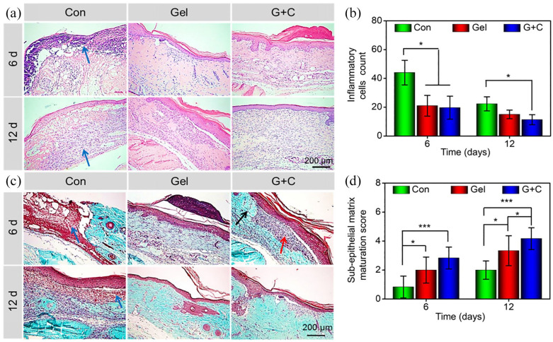
A diabetic foot ulcer (DFUs) is a state of prolonged chronic inflammation, which can result in amputation. Different from normal skin wounds, various commercially available dressings have not sufficiently improved the healing of DFUs. In this study, a novel self-healing hydrogel was prepared by in situ crosslinking of N-carboxyethyl chitosan (N-chitosan) and adipic acid dihydrazide (ADH) with hyaluronic acid-aldehyde (HA-ALD), to provide a moist and inflammatory relief environment to promote stem cell proliferation or secretion of growth factors, thus accelerating wound healing. The results demonstrated that this injectable and self-healing hydrogel has excellent swelling properties, stability, and mechanical properties. This biocompatible hydrogel stimulated secretion of growth factors from bone marrow mesenchymal stem cells (BM-MSCs) and regulated the inflammatory environment by inhibiting the expression of M1 macrophages and promoting the expression of M2 macrophages, resulting in granulation tissue formation, collagen deposition, nucleated cell proliferation, neovascularization, and enhanced diabetic wound healing. This study showed that N-chitosan/HA-ALD hydrogel could be used as a multifunctional injectable wound dressing to regulate chronic inflammation and provide an optimal environment for BM-MSCs to promote diabetic wound healing.

Keywords: BM-MSCs; Hydrogel; diabetic wound; inflammatory micro-environment.


Conflict of interest statement

Declaration of conflicting interests: The author(s) declared no potential conflicts of interest with respect to the research, authorship, and/or publication of this article.

Figures

Scheme 1.
Scheme 1.
A novel injectable hydrogel was prepared by in situ crosslinking of N-chitosan and ADH with HA-ALD, to provide a moist and inflammatory relief environment to promote BM-MSCs proliferation or secretion of growth factors, thus inducing collagen deposition and angiogenesis and accelerating wound healing.
Figure 1.
Figure 1.
(a) Optical images of the gelation progress. The mixture of N-chitosan and ADH was in the solution (sol) state at room temperature, and underwent sol–gel transition approximately 20 s after adding HA-ALD solution. (b) SEM image of the morphology and internal structure of the hydrogel. (c) Swelling properties of the hydrogel. (d) Degradation of the hydrogel.
Figure 2.
Figure 2.
(a) Representative images of the live/dead assay at 1 and 7 days on the hydrogel (Green: live cells, Red: dead cells). (b–d) The concentrations of the growth factors, TGF-β1, VEGF, and bFGF, secreted by BM-MSCs cultured in hydrogels for 1 and 3 days (Con and Gel represented the control and hydrogel groups, respectively. *p < 0.05, **p < 0.01).
Figure 3.
Figure 3.
Treatment effects of hydrogel + BMSCs on healing of DFUs. (a) Typical wound closure images of each group at different time points. (b) Time-course of wound changes. (c–e) The concentrations of the growth factors, TGF-β1, VEGF, and bFGF in wound tissue at 3 and 9 days after treatment. (Con, Gel, and G+C represented the control, hydrogel, and hydrogel + BM-MSCs groups, respectively. *p < 0.05, **p < 0.01, ***p < 0.001).
Figure 4.
Figure 4.
(a) H&E staining of the wound at 6 and 12 days after treatment (Blue arrows indicated necrotic tissue, foreign bodies, and a large number of inflammatory cells). (b) Inflammatory cell count in the wound area at 6 and 12 days after treatment. (c) Masson trichrome staining to detect new collagen fiber deposition in granulation tissue (Blue arrows indicated necrotic tissue, foreign bodies, and a large number of inflammatory cells; Red arrow indicated fibrous repair (mainly with collagen fibers), and black arrow indicated the original connective tissue of the dermis). At 12 days, the wound tissue in the hydrogel + BM-MSCs group was completely repaired, and was nearly identical in appearance to the surrounding tissue. (d) Sub-epithelial matrix maturation scores of the regeneration tissue at 6 and 12 days after treatment (Con, Gel, and G+C represented the control, hydrogel, and hydrogel + BM-MSCs groups, respectively. *p < 0.05, **p < 0.01, ***p < 0.001).
Figure 5.
Figure 5.
(a) Immunohistochemical staining of Ki67 in the wounds 6 and 12 days after treatment. (b) Quantitative analysis of mean density of Ki67 expression results in the wounds. (c) Immunohistochemical staining of CD31 expression in the wounds 6 and 12 days after treatment. (d) Neovascularization in the wounds (The black arrows represented the neovascularization) (Con, Gel, and G+C represented the control, hydrogel, and hydrogel + BM-MSCs groups, respectively. *p < 0.05, **p < 0.01).
Figure 6.
Figure 6.
Alleviation of chronic inflammation in DFUs. (a, b) Immunofluorescence staining and quantitative statistics of CD86-positive cells at wound sites. (c, d) Immunofluorescence staining and quantitative statistics of CD163-positive cells at wound sites (Con, Gel, and G+C represented the control, hydrogel, and hydrogel + BM-MSCs groups, respectively. *p < 0.05, **p < 0.01).
Figure 7.
Figure 7.
(a) H&E staining of the distribution of new sebaceous glands (purple arrow) and hair follicles (orange arrow) in the wound after 28 days of treatment. (b) Statistical analysis of new sebaceous glands. (c) Statistical analysis of new hair follicles (Con, Gel, and G+C represented the control, hydrogel, and hydrogel + BM-MSCs groups, respectively. *p < 0.05, n = 3).

Similar articles

Cited by

References

    1. Braffett BH, Gubitosi-Klug RA, Albers JW. Risk factors for diabetic peripheral neuropathy and cardiovascular autonomic neuropathy in the diabetes control and complications trial/epidemiology of diabetes interventions and complications (DCCT/EDIC) study. Diabetes 2020; 69(5): 1000–1010. - PMC - PubMed
    1. Whiting DR, Guariguata L, Weil C, et al. IDF diabetes atlas: global estimates of the prevalence of diabetes for 2011 and 2030. Diabetes Res Clin Pract 2011; 94(3): 311–321. - PubMed
    1. Brem H, Tomic-Canic M. Cellular and molecular basis of wound healing in diabetes. J Clin Invest 2007; 117(5): 1219–1222. - PMC - PubMed
    1. Moura LIF, Dias AMA, Carvalho E, et al. Recent advances on the development of wound dressings for diabetic foot ulcer treatment-A review. Acta Biomater 2013; 9(7): 7093–7114. - PubMed
    1. Zhao Y, Li Z, Song S, et al. Skin-inspired antibacterial conductive hydrogels for epidermal sensors and diabetic foot wound dressings. Adv Funct Mater 2019; 29(31): 1901474.

LinkOut - more resources