. 2020 Jul 25:18:372-381.
doi: 10.1016/j.omto.2020.07.007. eCollection 2020 Sep 25.

Polymethine Dye-Functionalized Nanoparticles for Targeting CML Stem Cells

Affiliations

Polymethine Dye-Functionalized Nanoparticles for Targeting CML Stem Cells

Philipp Ernst et al. Mol Ther Oncolytics. .

Abstract

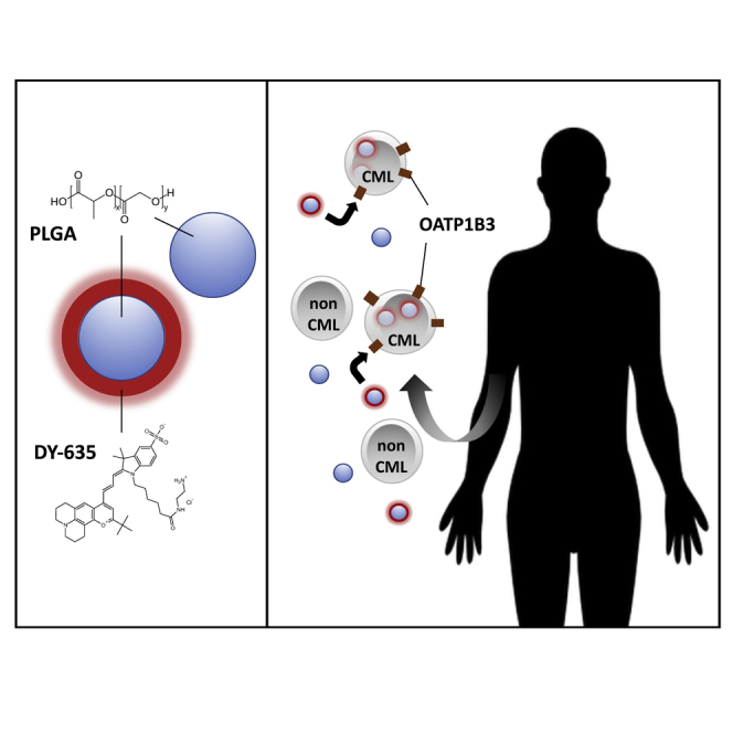
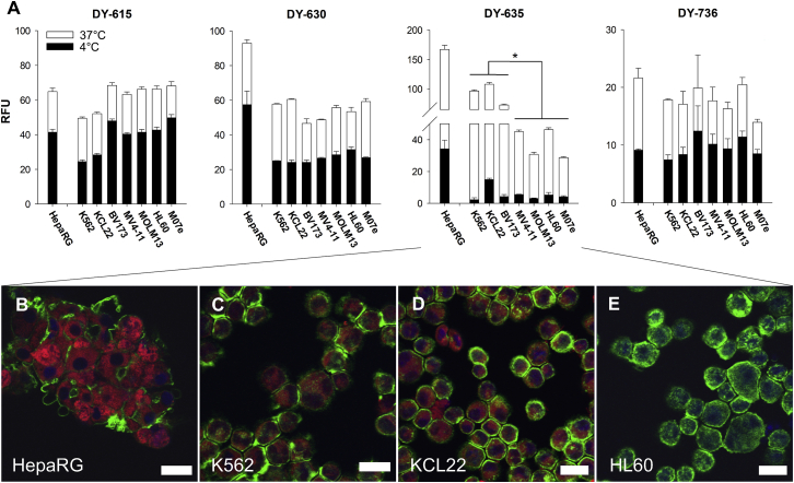
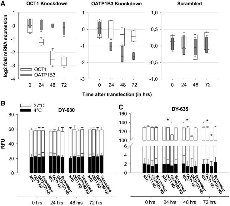
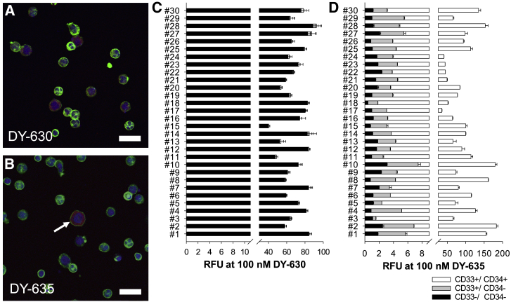
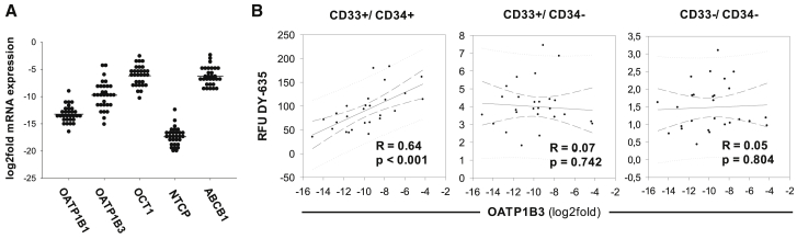
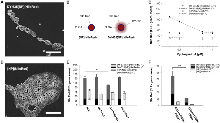
In chronic myelogenous leukemia (CML), treatment with tyrosine kinase inhibitors (TKI) is unable to eradicate leukemic stem cells (LSC). Polymethine dye-functionalized nanoparticles can be internalized by specific cell types using transmembrane carrier proteins. In this study we investigated the uptake behavior of various polymethine dyes on leukemia cell lines and searched for carrier proteins that guide dye transport using RNA interference. The results show that the uptake of DY-635 is dependent on organic anion transport protein 1B3 (OATP1B3) in CML cells and immature myeloid precursor cells of CML patients. In contrast to nonspecific poly(lactide-co-glycolic acid) (PLGA) nanoparticle constructs, DY-635-functionalization of nanoparticles led to an uptake in CML cells. Investigation of these nanoparticles on bone marrow of CML patients showed a preferred uptake in LSC. The transcription of OATP1B3 is known to be induced under hypoxic conditions via the hypoxia-inducing factor 1 alpha (HIF1α), thus also in the stem cells niche. Since these cells have the potential to repopulate the bone marrow after CML treatment discontinuation, eliminating them by means of drug-loaded DY-635-functionalized PLGA nanoparticles deployed as a selective delivery system to LSC is highly relevant to the ongoing search for curative treatment options for CML patients.

Keywords: CML; OATP1B3; OCT1; nanoparticles; polymethine dyes.


Figures

None
Graphical abstract
Figure 1
Figure 1
DY-635 Shows Different Uptake Behavior Dependent on the Cell Line (A) Flow cytometric determined relative fluorescence units (RFUs) after incubation of 100 nM DY-615, DY-630, DY-635, and Dy-736 with HepaRG cells, CML cell lines (K562, KCL22, BV173) and AML cell lines (MV4-11, MOLM13, M07e). Shown are mean values with their standard errors. ∗p < 0.05. (B–E) Laser scanning micrographs of HepaRG cells (B), K562 cells (C), KCL22 cells (D), and HL60 cells (E) after incubation with 100 nM DY-635 (red). Cytoskeleton staining with phalloidin Alexa 488 (green). Cell nucleus staining with DAPI-II (blue). Scale bars, 20 μm.
Figure 2
Figure 2
DY-635 Uptake Depends on the Presence of OATP1B3 (A) Quantitative real-time PCR of mRNA expression of OCT1 and OATP1B3 genes after siRNA knockdown in K562 cells. (B and C) Uptake of 100 nM DY-630 (B) and DY-635 (C) from both non-transfected K562 cells (NTCs) and K562 cells immediately before and 24, 48, and 72 h after transfection with OCT1, OATP1B3, or scrambled siRNA. KD, knockdown. RFU, relative fluorescence units. ∗p < 0.05.
Figure 3
Figure 3
Immature Precursor Cells of CML Patients Prefer DY-635 Uptake (A and B) Laser scanning microscopic analysis of mononuclear cells of a patient with newly diagnosed and untreated CML after incubation with 100 nM DY-630 (red in A) and 100 nM DY-635 (red in B). Immature precursor cell (arrow in B). Cytoskeleton (green, phalloidin Alexa 488). Nucleus (blue, DAPI-II). Scale bars, 20 μm. (C and D) Flow cytometric results from CD33/CD34 (black) and CD33+/CD34 (dark gray) and CD33+/CD34+ (white) cells from 30 patients with newly diagnosed and untreated CML after incubation with 100 nM DY-630 (C) and DY-635 (D). Shown are mean values with their standard deviation. RFU, relative fluorescence units.
Figure 4
Figure 4
OATP1B3 Expression of Immature Precursor Cells Is Associated with DY-635 Uptake in CML Patients (A) Density plots of the carrier protein expression in leukocytes from 30 CML patients. The mRNA expression of carrier proteins is shown as log2fold mRNA expression relative to the expression level of HepaRG cells. The horizontal line marks the mean value. (B) Pearson correlation of the RFUs of DY-635 of the cell fractions CD33+/CD34+, CD33+/CD34, and CD33/CD34 and the log2fold mRNA expression of OATP1B3. N = 30.
Figure 5
Figure 5
Functionalization of PLGA Nanoparticles with DY-635 Increases Nanoparticle Uptake in CML Cells with Stem Cell Signature (A) SEM of DY-635 functionalized PLGA nanoparticles DY-635[NP](NileRed). Scale bar, 2 μm. (B) Schematic illustration of non-functionalized nanoparticles [NP](NileRed) and DY-635[NP](NileRed) both labled with a Nile Red core. (C) Flow cytometric results of nanoparticle incubation of K562 cells with competitive antagonism of cyclosporin A. (D) SEM of [NP](NileRed). Scale bar, 2 μm. (E) Nanoparticle uptake of NTCs and 72 h after OCT1, OATP1B3, and scrambled siRNA transfection, respectively. Shown are mean values with their standard errors. KD, knockdown. (F) Flow cytometric results of nanoparticle incubation of CML cells with stem cell signature (CD34+/CD38/CD26+) and hematopoietic stem cells (CD34+/CD38/CD26). N = 5. ∗p < 0.05. ∗∗p < 0.01.

Similar articles

Cited by

References

    1. Eaves C., Udomsakdi C., Cashman J., Barnett M., Eaves A. The biology of normal and neoplastic stem cells in CML. Leuk. Lymphoma. 1993;11(Suppl 1):245–253. - PubMed
    1. Bhatia R., Holtz M., Niu N., Gray R., Snyder D.S., Sawyers C.L., Arber D.A., Slovak M.L., Forman S.J. Persistence of malignant hematopoietic progenitors in chronic myelogenous leukemia patients in complete cytogenetic remission following imatinib mesylate treatment. Blood. 2003;101:4701–4707. - PubMed
    1. Holyoake T., Jiang X., Eaves C., Eaves A. Isolation of a highly quiescent subpopulation of primitive leukemic cells in chronic myeloid leukemia. Blood. 1999;94:2056–2064. - PubMed
    1. Shah M., Bhatia R. Preservation of Quiescent Chronic Myelogenous Leukemia Stem Cells by the Bone Marrow Microenvironment. Adv. Exp. Med. Biol. 2018;1100:97–110. - PubMed
    1. Perl A., Carroll M. BCR-ABL kinase is dead; long live the CML stem cell. J. Clin. Invest. 2011;121:22–25. - PMC - PubMed

LinkOut - more resources