. 2020 Dec 1;35(5):391-404.
doi: 10.1093/mutage/geaa023.

DNA ligase I variants fail in the ligation of mutagenic repair intermediates with mismatches and oxidative DNA damage

Affiliations

DNA ligase I variants fail in the ligation of mutagenic repair intermediates with mismatches and oxidative DNA damage

Qun Tang et al. Mutagenesis. .

Abstract

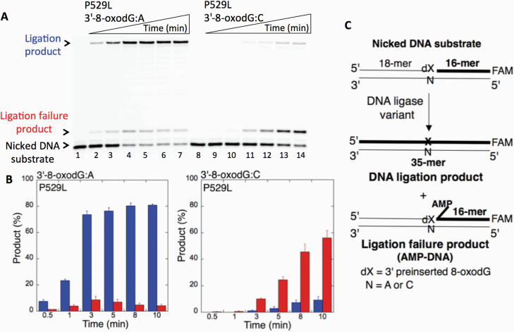
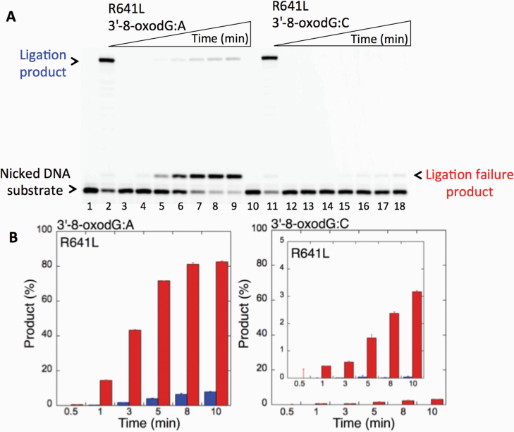
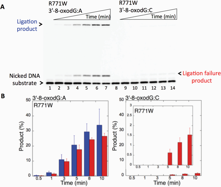
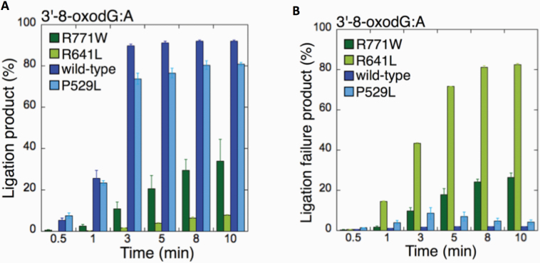
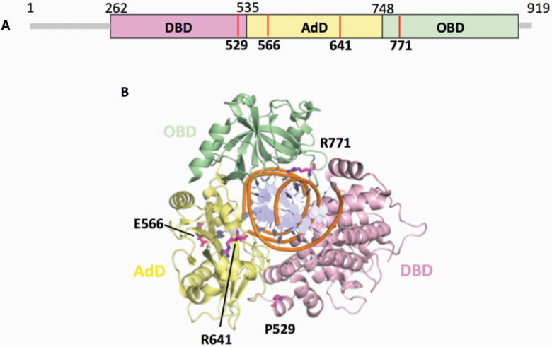
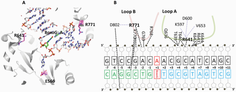
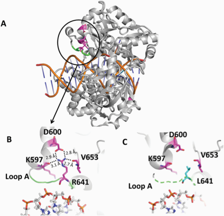
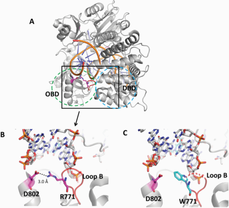
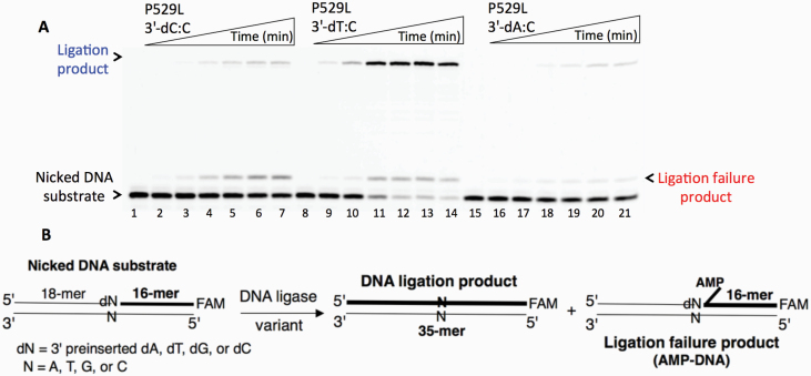
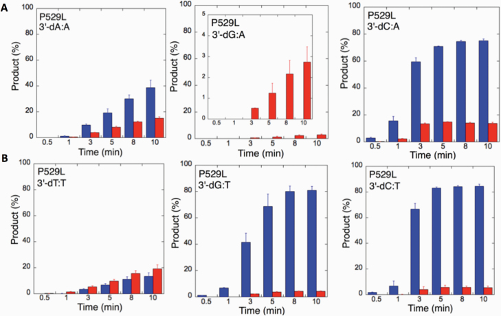
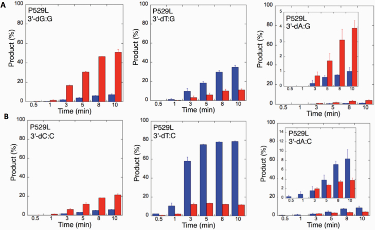
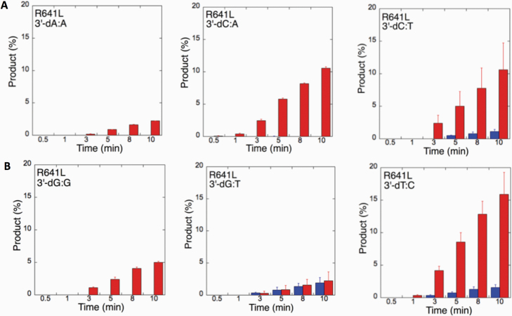
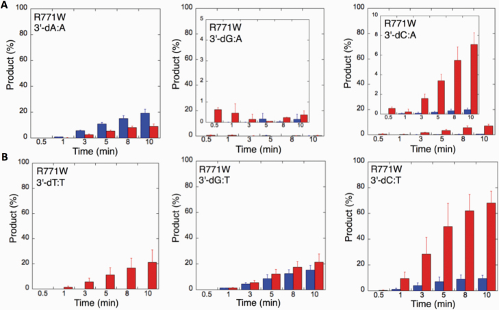
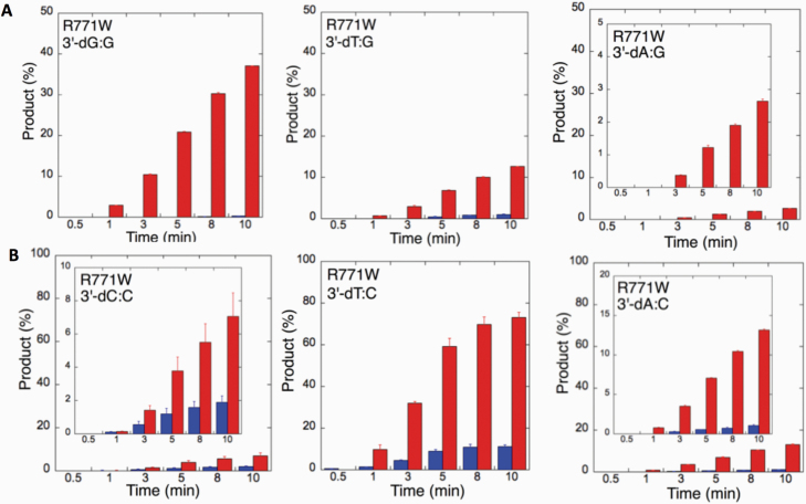
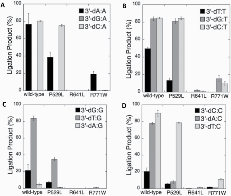
DNA ligase I (LIG1) joins DNA strand breaks during DNA replication and repair transactions and contributes to genome integrity. The mutations (P529L, E566K, R641L and R771W) in LIG1 gene are described in patients with LIG1-deficiency syndrome that exhibit immunodeficiency. LIG1 senses 3'-DNA ends with a mismatch or oxidative DNA base inserted by a repair DNA polymerase. However, the ligation efficiency of the LIG1 variants for DNA polymerase-promoted mutagenesis products with 3'-DNA mismatches or 8-oxo-2'-deoxyguanosine (8-oxodG) remains undefined. Here, we report that R641L and R771W fail in the ligation of nicked DNA with 3'-8-oxodG, leading to an accumulation of 5'-AMP-DNA intermediates in vitro. Moreover, we found that the presence of all possible 12 non-canonical base pairs variously impacts the ligation efficiency by P529L and R771W depending on the architecture at the DNA end, whereas E566K exhibits no activity against all substrates tested. Our results contribute to the understanding of the substrate specificity and mismatch discrimination of LIG1 for mutagenic repair intermediates and the effect of non-synonymous mutations on ligase fidelity.


Figures

Figure 1.
Figure 1.
Ligation of the repair intermediate with 3’-8-oxodG by LIG1 variant P529L. (A) Lanes 1 and 8 are the negative enzyme controls of the nicked DNA substrates with 3’-8-oxodG opposite templates A and C, respectively. Lanes 2–7 and 9–14 show the reaction products obtained for 3’-8-oxodG:A and 3’-8-oxodG:C substrates, respectively, at Time points 0.5, 1, 3, 5, 8 and 10 min. (B) The graphs show the time-dependent changes in the ligation (blue) and ligation failure (red) products, and the data are presented as the averages from three independent experiments ± SDs. (C) Illustrations of the nicked DNA substrate with 3’-8-oxodG and the products observed in the ligation reaction.
Figure 2.
Figure 2.
Ligation of the repair intermediate with 3’-8-oxodG by LIG1 variant R641L. (A) Lanes 1 and 10 are the negative enzyme controls of the nicked DNA substrates with 3’-dT opposite template A and 3’-dC opposite template G, respectively. Lanes 2 and 11 show the reaction products obtained for 3’-dT:A and 3’-dC:G, respectively. Lanes 3 and 12 are the negative enzyme controls of the nicked DNA substrates with 3’-8-oxodG opposite templates A and C, respectively. Lanes 4–9 and 13–18 show the reaction products obtained for 3’-8-oxodG:A and 3’-8-oxodG:C substrates, respectively, at Time points 0.5, 1, 3, 5, 8 and 10 min. (B) The graphs show the time-dependent changes in the ligation (blue) and ligation failure (red) products, and the data are presented as the averages from three independent experiments ± SDs.
Figure 3.
Figure 3.
Ligation of the repair intermediate with 3’-8-oxodG by LIG1 variant R771W. (A) Lanes 1 and 8 are the negative enzyme controls of the nicked DNA substrates with 3’-8-oxodG opposite templates A and C, respectively. Lanes 2–7 and 9–14 show the reaction products obtained for 3’-8-oxodG:A and 3’-8-oxodG:C substrates, respectively, at Time points 0.5, 1, 3, 5, 8 and 10 min. (B) The graphs show the time-dependent changes in the ligation (blue) and ligation failure (red) products, and the data are presented as the averages from three independent experiments ± SDs.
Figure 4.
Figure 4.
Comparisons for the ligation of 3’-8-oxodG:A substrate by the wild-type and LIG1 variants. (A, B) The graphs show the time-dependent changes in the ligation (A) and ligation failure (B) products for the wild-type and LIG1 variants P529L, R641L and R771W. The data are presented as the averages from three independent experiments ± SDs.
Figure 5.
Figure 5.
Domain architecture of LIG1 with the disease mutations. (A) Schematic view showing the domain composition of human LIG1, including the N-terminal domain (amino acids 1–261, gray), and the catalytic core (amino acids 262–919) consisting of the DBD (pink), AdD (yellow) and OBD (green). (B) LIG1 (cartoon) in complex with an adenylated nicked DNA complex (stick, orange). The amino acid residues (magenta), P529, E566, R641 and R771, that are mutated in LIG1-deficiency disease are shown as sticks.
Figure 6.
Figure 6.
Architecture of LIG1 showing the positions of the disease-associated residues in complex with the nicked DNA containing 8-oxoG:A. (A) Ribbon diagram shows LIG1 (grey) encircling the nicked DNA (sticks) with 8-oxoG opposite template A (green). E566, R641 and R771 (magenta) are represented by sticks. (B) Nucleotide-residue contact map showing individual LIG1 residues interactions that are close proximity to R641 (loop A, green) and R771 (loop B, red) with the nicked DNA containing 8-oxoG:A.
Figure 7.
Figure 7.
Structure model of LIG1 with R641L identified in LIG1 deficiency syndrome. (A) Ribbon diagram showing LIG1 encircling a nicked DNA duplex (orange). Arg641 (magenta) shown as sticks is located in Loop A (green). (B) Arg641 (R641) interacts with K597, D600 and V653, and (C) the replacement of Arg641 with Leu (L641) disrupts the interaction and destabilises Loop A (shown in dotted green line). All residues are shown as sticks. Positive and negative potentials are shown in blue and red, respectively.
Figure 8.
Figure 8.
Structure model of LIG1 with R771W identified in LIG1 deficiency syndrome. (A) Ribbon diagram showing LIG1 encircling a nicked DNA duplex (orange). Arg771 (magenta) shown as sticks is located in Loop B (red) that resides in the OBD domain (green dotted circle) and interacts with the DBD (blue dotted circle). (B) Arg771 (R771) interacts with D802 (magenta) to stabilise the loop B, and (C) replacement of Arg771 with Trp (cyan, W771) disrupts this interaction and destabilises Loop B (shown in dotted red line). All residues are shown as sticks. Positive and negative potentials are shown in blue and red, respectively.
Figure 9.
Figure 9.
Ligation of the repair intermediate with 3’-pre-inserted mismatches opposite template C by LIG1 variant P529L. (A) Lanes 1, 8 and 15 are the negative enzyme controls of the nicked DNA substrates with 3’-dC:C, 3’-dT:C and 3’-dA:C, respectively. Lanes 2–7, 9–14 and 16–21 show the reaction products obtained at time points 0.5, 1, 3, 5, 8 and 10 min. (B) Illustrations of the nicked DNA substrate with 3’-mismatches and the products observed in the ligation reaction.
Figure 10.
Figure 10.
Specificity of LIG1 variant P529L for the ligation of repair intermediates with 3’-pre-inserted mismatches opposite templates A and T. (A,B) The graphs show the time-dependent changes in the ligation (blue) and ligation failure (red) products for 3’-pre-inserted mismatches opposite templates A (A) and T (B). The data are presented as the averages from three independent experiments ± SDs. The gel images are presented in Supplementary Figure 5.
Figure 11.
Figure 11.
Specificity of LIG1 variant P529L for the ligation of repair intermediates with 3’-pre-inserted mismatches opposite templates G and C. (A, B) The graphs show the time-dependent changes in the ligation (blue) and ligation failure (red) products for 3’-pre-inserted mismatches opposite templates G (A) and C (B). The data are presented as the averages from three independent experiments ± SDs. The gel images are presented in Figure 9 and Supplementary Figure 6.
Figure 12.
Figure 12.
Specificity of LIG1 variant R641L for the ligation of repair intermediates with 3’-pre-inserted mismatches. (A, B) The graphs show the time-dependent changes in the ligation (blue) and ligation failure (red) products. The data are presented as the averages from three independent experiments ± SDs. The gel images are presented in Supplementary Figures 7 and 8.
Figure 13.
Figure 13.
Specificity of LIG1 variant R771W for the ligation of repair intermediates with 3’-pre-inserted mismatches opposite templates A and T. (A, B) The graphs show the time-dependent changes in the ligation (blue) and ligation failure (red) products for 3’-pre-inserted mismatches opposite templates A (A) and T (B). The data are presented as the averages from three independent experiments ± SDs. The gel images are presented in Supplementary Figure 9.
Figure 14.
Figure 14.
Specificity of R771W for the ligation of repair intermediates with 3’-pre-inserted mismatches opposite templates G and C. (A, B) The graphs show the time-dependent changes in the ligation (blue) and ligation failure (red) products for 3’-pre-inserted mismatches opposite templates G (A) and C (B). The data are presented as the averages from three independent experiments ± SDs. The gel images are presented in Supplementary Figure 10.
Figure 15.
Figure 15.
Comparisons for the ligation of 3’-pre-inserted mismatch-containing substrates by the wild-type and LIG1 variants. (A–D) The graphs show the time-dependent changes in the amount of ligation products, and the data are presented as the averages from three independent experiments ± SDs.

Similar articles

Cited by

References

    1. De Bont R and van Larebeke N (2004) Endogenous DNA damage in humans: a review of quantitative data. Mutagenesis, 19, 169–185. - PubMed
    1. Singh R, Kaur B, Kalina I, et al. (2007) Effects of environmental air pollution on endogenous oxidative DNA damage in humans. Mutat. Res., 620, 71–82. - PubMed
    1. Nelson B C and Dizdaroglu M (2020) Implications of DNA damage and DNA repair on human diseases. Mutagenesis, 35, 1–3. - PMC - PubMed
    1. Chatterjee N and Walker G C (2017) Mechanisms of DNA damage, repair, and mutagenesis. Environ. Mol. Mutagen., 58, 235–263. - PMC - PubMed
    1. Ellenberger T and Tomkinson A E (2008) Eukaryotic DNA ligases: structural and functional insights. Annu. Rev. Biochem., 77, 313–338. - PMC - PubMed

Publication types

MeSH terms