

Advances in optical gastrointestinal endoscopy: a technical review
- PMID: 32915503
- PMCID: PMC8486567
- DOI: 10.1002/1878-0261.12792
Advances in optical gastrointestinal endoscopy: a technical review
Abstract
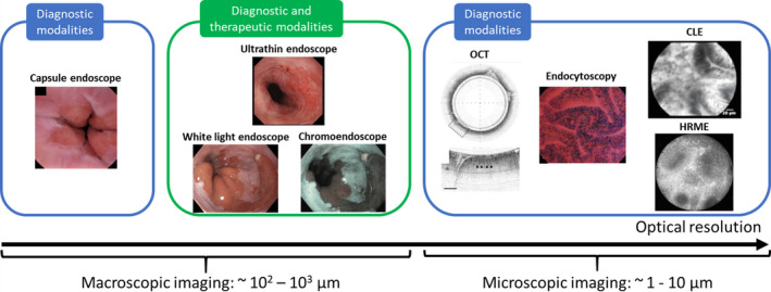
Optical endoscopy is the primary diagnostic and therapeutic tool for management of gastrointestinal (GI) malignancies. Most GI neoplasms arise from precancerous lesions; thus, technical innovations to improve detection and diagnosis of precancerous lesions and early cancers play a pivotal role in improving outcomes. Over the last few decades, the field of GI endoscopy has witnessed enormous and focused efforts to develop and translate accurate, user-friendly, and minimally invasive optical imaging modalities. From a technical point of view, a wide range of novel optical techniques is now available to probe different aspects of light-tissue interaction at macroscopic and microscopic scales, complementing white light endoscopy. Most of these new modalities have been successfully validated and translated to routine clinical practice. Herein, we provide a technical review of the current status of existing and promising new optical endoscopic imaging technologies for GI cancer screening and surveillance. We summarize the underlying principles of light-tissue interaction, the imaging performance at different scales, and highlight what is known about clinical applicability and effectiveness. Furthermore, we discuss recent discovery and translation of novel molecular probes that have shown promise to augment endoscopists' ability to diagnose GI lesions with high specificity. We also review and discuss the role and potential clinical integration of artificial intelligence-based algorithms to provide decision support in real time. Finally, we provide perspectives on future technology development and its potential to transform endoscopic GI cancer detection and diagnosis.
Keywords: gastrointestinal tract; machine learning; molecular probe; optical endoscopy.
© 2020 The Authors. Published by FEBS Press and John Wiley & Sons Ltd.
Conflict of interest statement
The authors declare no conflict of interest.
Figures
References
-
- Bray F, Ferlay J, Soerjomataram I, Siegel RL, Torre LA & Jemal A (2018) Global cancer statistics 2018: GLOBOCAN estimates of incidence and mortality worldwide for 36 cancers in 185 countries. CA Cancer J Clin 68, 394–424. - PubMed
-
- Dawsey SM, Fleischer DE, Wang G‐Q, Zhou B, Kidwell JA, Lu N, Lewin KJ, Roth MJ, Tio TL & Taylor PR (1998) Mucosal iodine staining improves endoscopic visualization of squamous dysplasia and squamous cell carcinoma of the esophagus in linxian, china. Cancer 83, 220–231. - PubMed
Publication types
MeSH terms
Grants and funding
LinkOut - more resources
Full Text Sources
