Gallbladder polyps: Correlation of size and clinicopathologic characteristics based on updated definitions
- PMID: 32915805
- PMCID: PMC7485812
- DOI: 10.1371/journal.pone.0237979
Gallbladder polyps: Correlation of size and clinicopathologic characteristics based on updated definitions
Erratum in
-
Correction: Gallbladder polyps: Correlation of size and clinicopathologic characteristics based on updated definitions.PLoS One. 2024 Jul 5;19(7):e0306997. doi: 10.1371/journal.pone.0306997. eCollection 2024. PLoS One. 2024. PMID: 38968267 Free PMC article.
Abstract
Background: Different perspectives exist regarding the clinicopathologic characteristics, biology and management of gallbladder polyps. Size is often used as the surrogate evidence of polyp behavior and size of ≥1cm is widely used as cholecystectomy indication. Most studies on this issue are based on the pathologic correlation of polyps clinically selected for resection, whereas, the data regarding the nature of polypoid lesions from pathology perspective -regardless of the cholecystectomy indication- is highly limited.
Methods: In this study, 4231 gallbladders -606 of which had gallbladder carcinoma- were reviewed carefully pathologically by the authors for polyps (defined as ≥2 mm). Separately, the cases that were diagnosed as "gallbladder polyps" in the surgical pathology databases were retrieved.
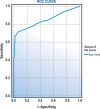
Results: 643 polyps identified accordingly were re-evaluated histopathologically. Mean age of all patients was 55 years (range: 20-94); mean polyp size was 9 mm. Among these 643 polyps, 223 (34.6%) were neoplastic: I. Non-neoplastic polyps (n = 420; 65.4%) were smaller (mean: 4.1 mm), occurred in younger patients (mean: 52 years). This group consisted of fibromyoglandular polyps (n = 196) per the updated classification, cholesterol polyps (n = 166), polypoid pyloric gland metaplasia (n = 41) and inflammatory polyps (n = 17). II. Neoplastic polyps were larger (mean: 21 mm), detected in older patients (mean: 61 years) and consisted of intra-cholecystic neoplasms (WHO's "adenomas" and "intracholecystic papillary neoplasms", ≥1 cm; n = 120), their "incipient" version (<1 cm) (n = 44), polypoid invasive carcinomas (n = 26) and non-neoplastic polyps with incidental dysplastic changes (n = 33). In terms of size cut-off correlations, overall, only 27% of polyps were ≥1 cm, 90% of which were neoplastic. All (except for one) ≥2 cm were neoplastic. However, 14% of polyps <1 cm were also neoplastic. Positive predictive value of ≥1 cm cut-off -which is widely used for cholecystectomy indication-, was 94.3% and negative predictive value was 85%.
Conclusions: Approximately a third of polypoid lesions in the cholecystectomies (regardless of the indication) prove to be neoplastic. The vast majority of (90%) of polyps ≥1 cm and virtually all of those ≥2 cm are neoplastic confirming the current impression that polyps ≥1 cm ought to be removed. However, this study also illustrates that 30% of the neoplastic polyps are <1 cm and therefore small polyps should also be closely watched, especially in older patients.
Conflict of interest statement
The authors of this study have no competing interests to declare.
Figures


References
-
- Wiles R, Thoeni RF, Barbu ST, Vashist YK, Rafaelsen SR, Dewhurst C, et al. Management and follow-up of gallbladder polyps: Joint guidelines between the European Society of Gastrointestinal and Abdominal Radiology (ESGAR), European Association for Endoscopic Surgery and other Interventional Techniques (EAES), International Socie. Eur Radiol. 2017;27: 3856–3866. 10.1007/s00330-017-4742-y - DOI - PMC - PubMed
MeSH terms
Grants and funding
LinkOut - more resources
Full Text Sources
Medical

