Review
. 2020 Nov 23:54:487-510.
doi: 10.1146/annurev-genet-041720-093403. Epub 2020 Sep 11.

Mosaicism in Human Health and Disease

Affiliations
Review

Mosaicism in Human Health and Disease

Jeremy Thorpe et al. Annu Rev Genet. .

Abstract

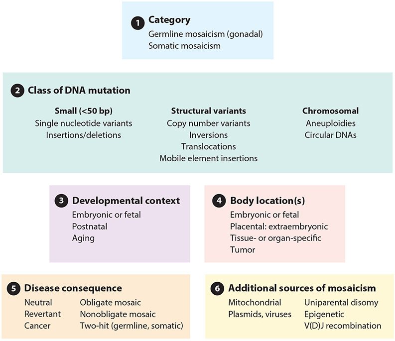
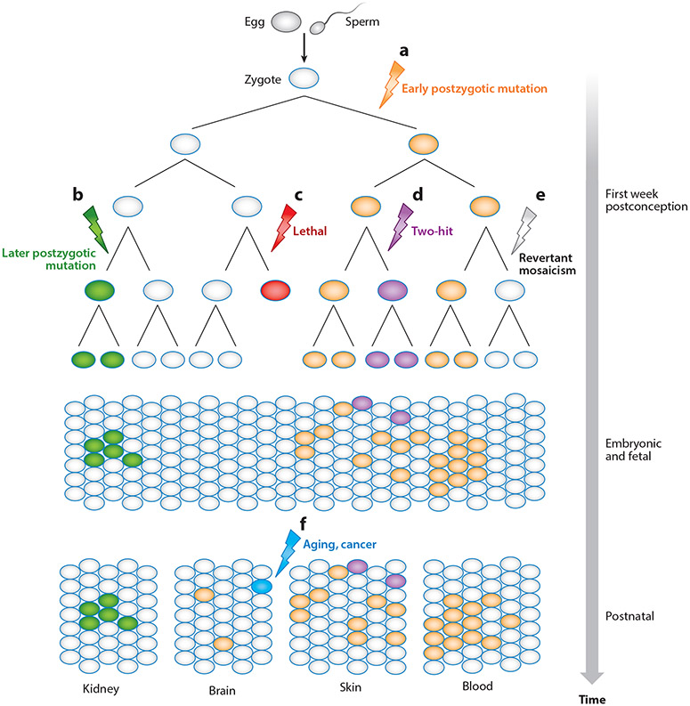
Mosaicism refers to the occurrence of two or more genomes in an individual derived from a single zygote. Germline mosaicism is a mutation that is limited to the gonads and can be transmitted to offspring. Somatic mosaicism is a postzygotic mutation that occurs in the soma, and it may occur at any developmental stage or in adult tissues. Mosaic variation may be classified in six ways: (a) germline or somatic origin, (b) class of DNA mutation (ranging in scale from single base pairs to multiple chromosomes), (c) developmental context, (d) body location(s), (e) functional consequence (including deleterious, neutral, or advantageous), and (f) additional sources of mosaicism, including mitochondrial heteroplasmy, exogenous DNA sources such as vectors, and epigenetic changes such as imprinting and X-chromosome inactivation. Technological advances, including single-cell and other next-generation sequencing, have facilitated improved sensitivity and specificity to detect mosaicism in a variety of biological contexts.

Keywords: germline mutation; mosaic aneuploidy; mosaicism; somatic mutation.


Figures

Figure 1
Figure 1
Classification of mosaic variation. We classify mosaic variation as: (a) germline or somatic origin, (b) class of DNA mutation (ranging in scale from single base pairs to multiple chromosomes), (c) developmental context, (d) body location(s), (e) disease consequence, and (f) additional sources of mosaicism.
Figure 2
Figure 2
Occurrence of somatic mosaicism across embryonic and fetal development, including development from the morula (16-cell stage) and blastocyst. The embryo forms from the inner cell mass of the blastocyst, while the placenta develops from the outer cell mass (from the trophoblasts at the exterior of the blastocyst). Mutations are indicated with a lightning bolt symbol, (a) Euploid (wild type) development. A normal karyotype (46,XX for a female in this example) is indicated, (b Germline mosaicism. Mutation can occur in the germline (e.g., germ cell precursors) of an embryo. Mosaic mutation is limited to germ cells that may later be transmitted to offspring as inherited germline variations. The individual depicted here, harboring the germ cell mosaic, may eventually become a parent who is phenotypieally normal, (c) Postzygotic mosaic mutations. Whether involving aneuploidy (whole chromosome gains or losses) or single nucleotide variants, postzygotic mosaic mutations can manifest through the entire body of an individual, particularly if the mutation occurs early in development, or be constrained to particular regions or organs, as indicated here. Trisomies that are ordinarily lethal (trisomies 1–12, 14–17, 19–20, and 22) may persist in the mosaic state with variable clinical phenotypes, (d) Confined placental mosaicism. Mosaic mutations can occur solely in the placenta while the fetus has a normal karyotype. A postzygotic, somatic mutation may lead to a cell and its daughter cells having mosaicism (such as mos 46,XX/47,XX+21 for mosaic trisomy 21) in a subset of the placental cells. Chorionic villus sampling can indicate trisomy 21. Determining whether the fetus is euploid or trisomic requires a separate test, such as amniocentesis, (e) Uniparental disomy. A trisomy (such as 47,XX+15) can arise at an early embryonic stage, persisting as a mosaic in the placenta (mos 46,XX/47,XX+15). In the embryo, trisomic rescue may occur in which a third copy of the chromosome is deleted from the cell. Rescue produces disomy and a euploid state (46,XX) or disomy in which both copies of the chromosome derive from one parent [e.g., maternal uniparental disomy (UPD mat)]. Uniparental heterodisomy refers to a meiosis I error, and both copies of the chromosome are from one parent but from different homologs. Uniparental isodisomy results from a meiosis II error or occurs through postzygotic duplication (as shown here). In this example, the child is susceptible to developing Prader-Willi syndrome [Online Mendelian Inheritance in Man (OMIM) 176270], an imprinting disorder due to loss of the paternal copy of chromosome 15q11.2–q13. This figure was derived in part from a public domain image from the Human Placenta Project (https://www.nichd.nih.gov/sites/default/files/inline-images/HPP-placental-development.png) at the Eunice Kennedy Shriver National Institute of Child Health and Human Development (National Institutes of Health).
Figure 3
Figure 3
Inference of germline mosaicism by pedigree analysis. In this pedigree, individuals III.4 and III. 5 present with an inherited autosomal dominant trait. Nevertheless, the father, II.2, is healthy. Individuals III.4 and III.5 are born from two different mothers, suggesting that the father, who does not show a clinical phenotype, is a gonadal mosaic for the mutation carried by offspring III.4 and III. 5. The genetic risk of transmitting the autosomal dominant condition to generation III subjects differs depending on the proportion of mutated sperm and the disease mechanism (autosomal dominant in this example). Siblings III.2 and III. 3 are healthy, having inherited the normal paternal allele, and are at no risk of transmitting the disease to their children. Subjects III.4 and III. 5 carry the germline mutation inherited by their father and have the mutation in both their germ and somatic cells. As for any autosomal dominant trait, there is a 50% probability of transmitting the mutant allele to their children (e.g., individual IV.3 is affected but IV.4 is not). In males, it is possible to confirm the mutation as germline mosaic by searching for it in DNA from sperm and somatic cells, for example, in peripheral blood lymphocytes. The mutation is germline mosaic if it is detected in sperm but not in lymphocyte DNA. The arrow indicates individual III. 5 who is the proband in this pedigree.
Figure 4
Figure 4
Somatic mosaicism across development and body regions and schematic representation of somatic mosaicism through cellular development from early life (top) to adulthood (bottom). Lightning bolts represent the occurrence of a somatic mosaic mutation, (a) An early postzygotic mutation affects one of two parental alleles. If the affected cell can divide and proliferate, it is likely to affect a relatively large part of the body (see orange cells, bottom), (b) A later-occurring somatic mutation may result in fewer affected cells, for example, restricted, organ-specific mosaicism, (c) Lethal somatic mosaic mutation can occur, (d) Additional, independent mosaic mutations can occur, (e) Revertant mosaicism occurs when a cell retrogresses the mosaic mutation to wild type, (f) Somatic mosaic mutations may occur at any time in development, from postconception to old age. An acquired somatic mutation is indicated, such as may occur as part of the aging process or in response to a mutagen.

References

    1. Ajay SS, Parker SC, Abaan HO, Fajardo KV, Margulies EH. 2011. Accurate and comprehensive sequencing of personal genomes. Genome Res. 21:1498–505 - PMC - PubMed
    1. Alexandrov LB, Kim J, Haradhvala NJ, Huang MN, Tian Ng AW, et al.2020. The repertoire of mutational signatures in human cancer. Nature 578:94–101 - PMC - PubMed
    1. Antonarakis SE. 2017. Down syndrome and the complexity of genome dosage imbalance. Nat. Rev. Genet 18:147–63 - PubMed
    1. Ardui S, Ameur A, Vermeesch JR, Hestand MS. 2018. Single molecule real-time (SMRT) sequencing comes of age: applications and utilities for medical diagnostics. Nucleic Acids Res. 46:2159–68 - PMC - PubMed
    1. Arvey A, Hermann A, Hsia CC, Ie E, Freund Y, McGinnis W. 2010. Minimizing off-target signals in RNA fluorescent in situ hybridization. Nucleic Acids Res. 38:e115. - PMC - PubMed

Publication types

LinkOut - more resources