. 2020 Dec 1:518:111025.
doi: 10.1016/j.mce.2020.111025. Epub 2020 Sep 8.

Biological behaviors of mutant proinsulin contribute to the phenotypic spectrum of diabetes associated with insulin gene mutations

Affiliations

Biological behaviors of mutant proinsulin contribute to the phenotypic spectrum of diabetes associated with insulin gene mutations

Heting Wang et al. Mol Cell Endocrinol. .

Abstract

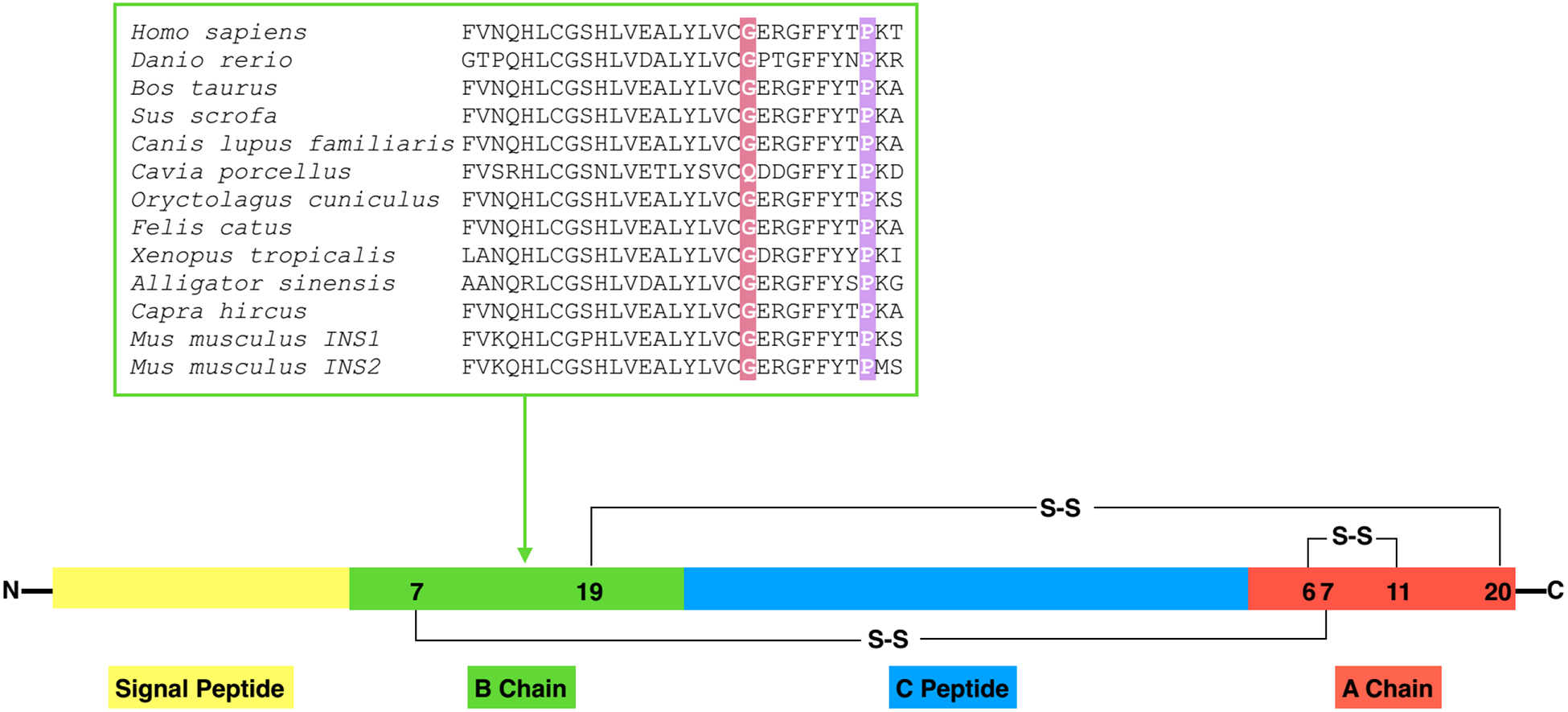
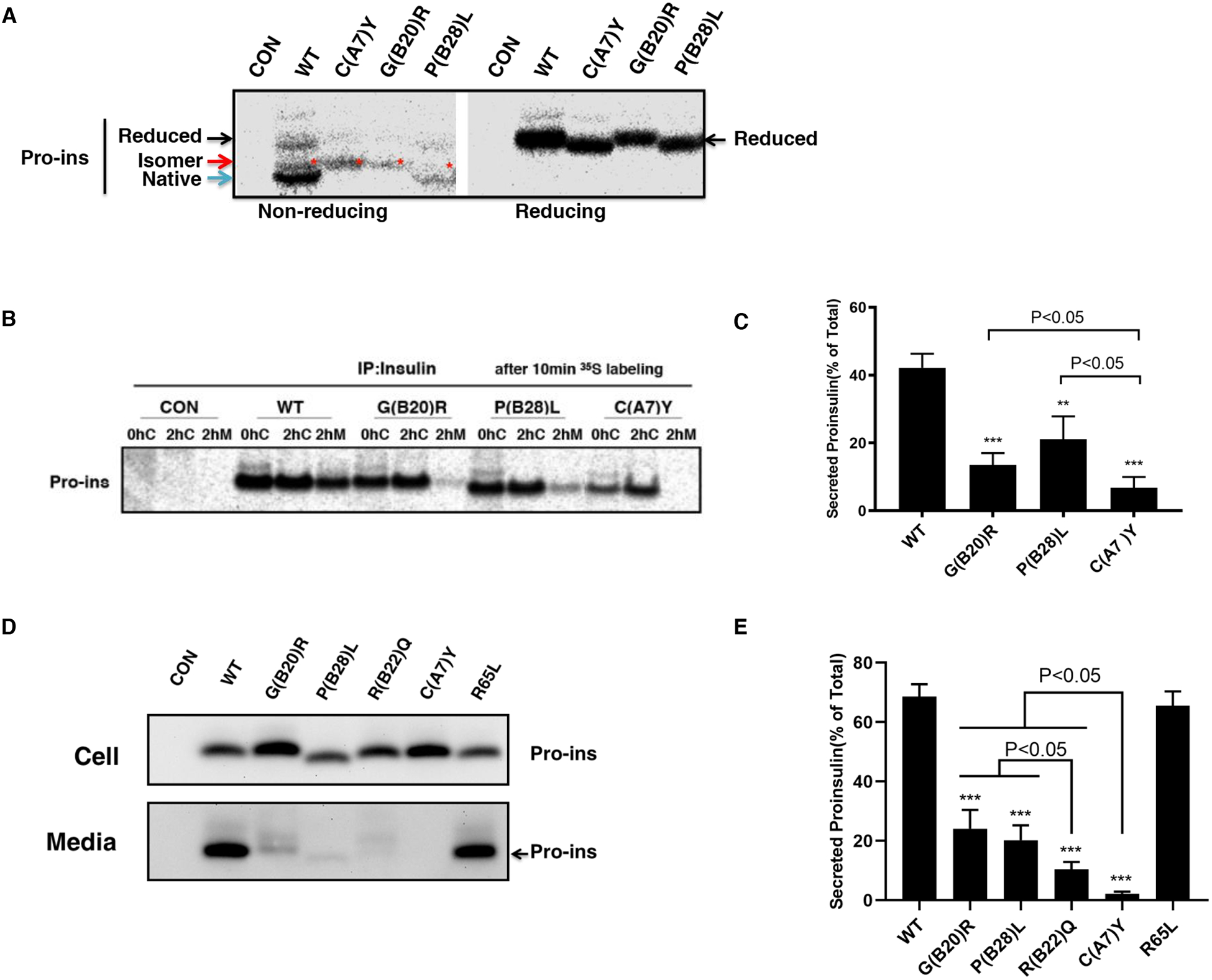
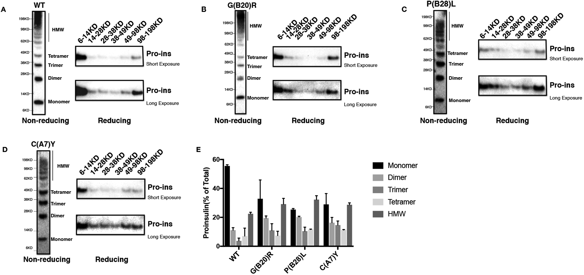
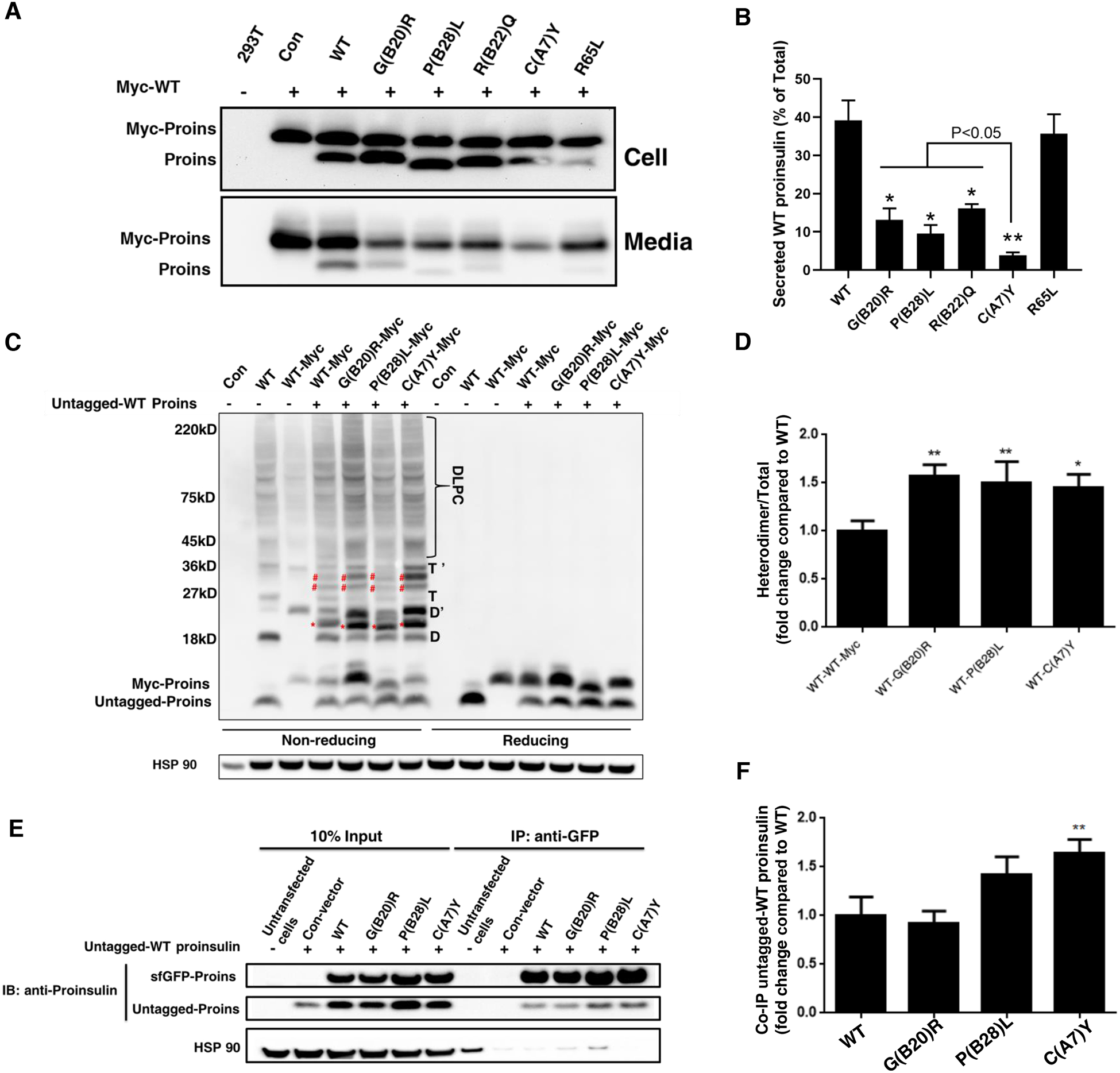
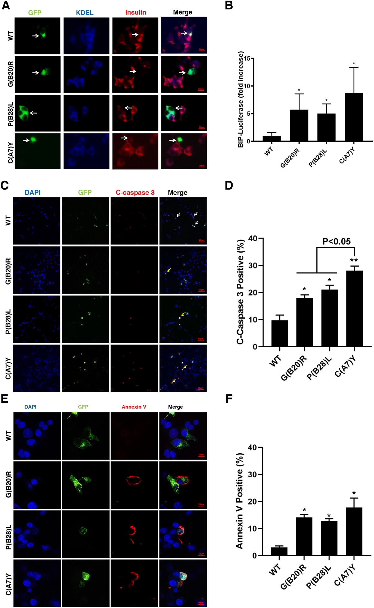
Insulin gene mutation is the second most common cause of neonatal diabetes (NDM). It is also one of the genes involved in maturity-onset diabetes of the young (MODY). We aim to investigate molecular behaviors of different INS gene variants that may correlate with the clinical spectrum of diabetes phenotypes. In this study, we concentrated on two previously uncharacterized MODY-causing mutants, proinsulin-p.Gly44Arg [G(B20)R] and p.Pro52Leu [P(B28)L] (a novel mutant identified in one French family), and an NDM causing proinsulin-p.(Cys96Tyr) [C(A7)Y]. We find that these proinsulin mutants exhibit impaired oxidative folding in the endoplasmic reticulum (ER) with blocked ER export, ER stress, and apoptosis. Importantly, the proinsulin mutants formed abnormal intermolecular disulfide bonds that not only involved the mutant proinsulin, but also the co-expressed WT-proinsulin, forming misfolded disulfide-linked proinsulin complexes. This impaired the intracellular trafficking of WT-proinsulin and limited the production of bioactive mature insulin. Notably, although all three mutants presented with similar defects in folding, trafficking, and dominant negative behavior, the degrees of these defects appeared to be different. Specifically, compared to MODY mutants G(B20)R and P(B28)L that partially affected folding and trafficking of co-expressed WT-proinsulin, the NDM mutant C(A7)Y resulted in an almost complete blockade of the ER export of WT-proinsulin, decreasing insulin production, inducing more severe ER stress and apoptosis. We thus demonstrate that differences in cell biological behaviors among different proinsulin mutants correlate with the spectrum of diabetes phenotypes caused by the different INS gene mutations.

Keywords: Dominant negative effect; ER stress; Insulin gene mutations; Maturity onset diabetes of the young; Neonatal diabetes mellitus; Proinsulin misfolding.


Conflict of interest statement

Declaration of competing interest

None.

Figures

Fig. 1.
Fig. 1.. Highly conserved amino acids B20 and B28 in different species.
Structure of preproinsulin: The signal peptide (SP, yellow), insulin B chain (green), C peptide (blue), insulin A chain (red), S-S indicate the B19-A20, A6-A11, B7-A7 three different disulfide bonds. B chain alignment between human INS and orthologs. Amino acids B20 and B28 were marked with red and purple, respectively.
Fig. 2.
Fig. 2.. INS variants impair proinsulin oxidative folding and ER export.
A. 293T cells were transfected with plasmids encoding wild-type (WT), or mutants C(A7)Y, G(B20)R, P(B28)L. At 48 h post-transfection, the cells were labeled with 35S-Met/Cys for 10 min. The newly synthesized proinsulin were precipitated with anti-insulin and analyzed by Tris–tricine–urea–SDS-PAGE under both non-reducing or reducing conditions. Reduced forms proinsulin marked by the black arrow, native forms marked by the blue arrow, disulfide isomers marked by the red arrow and star. B. 293T cells were transfected as Fig. 2A and pulse-labeled at 48 h with 35S-Met/Cys for 10 min followed by 0 or 2 h chase. Both cell lysates harvested after 0h (0hC) or 2h (2hC) chase and chase media (2hM) were immunoprecipitated with the anti-insulin and analyzed in 4–12% NuPage gel under reducing conditions with autoradiography. C. The secretion efficiency of WT or mutant proinsulin from at least three independent experiments shown in Fig. 2B was quantified using ImageJ. The results were shown as mean±SD, ** p <0.01 and *** p <0.001 comparing to WT (ANOVA test). D. 293T cells were transfected with plasmids encoding WT or mutant proinsulin as indicated. At 24 h post-transfection, the culture media were changed. After additional 24 hour incubation, the media were collected and cells were lysed. Both media and lysates were subjected to western blotting using anti-proinsulin antibody. E. The secretion efficiency of WT or mutant proinsulin under steady state from at least three independent experiments shown in Fig. 2D was quantified using ImageJ. The results were shown as mean±SD, *** p <0.001 comparing to WT (ANOVA test).
Fig. 3.
Fig. 3.. Proinsulin mutants form misfolded disulfide-linked proinsulin complexes (DLPC) in the ER.
293T cells were transfected with Myc-tagged plasmids encoding WT or mutant proinsulin. At 48 h post-transfection, cell lysates were resolved in 4–12% NuPage under non-reducing condition (left panel). The gels were cut into 6 pieces corresponding to the molecular weight, then boiled in the sample buffer containing 100mM DTT followed by resolved again in 4–12% NuPage. The gels were transferred to nitrocellulose following by blotting with anti-proinsulin antibody. The same procedure was processed both for WT (A), G(B20)R (B), P(B28)L (C), and C(A7)Y (D). E. The 2-DE assay shown in Fig. 3A-D from at least three independent experiments was quantified using ImageJ. The percentages of fully reduced proinsulin monomer(6–14KD), dimer(14–28KD), trimer(28–38KD), tetramer(38–49KD) and high molecular weight (HMW) complexes (49–198KD) in total proinsulin molecules were calculated and shown as mean±SD.
Fig.4.
Fig.4.. Proinsulin mutants interact with co-expressed WT-proinsulin and impair the ER export of WT-proinsulin.
A-B. 293T cells were co-transfected with Myc-tagged WT-proinsulin (upper bands) and untagged WT-proinsulin or mutants (lower bands) as indicated. The secretion of Myc-tagged WT-proinsulin in the presence of untagged WT-proinsulin or mutants under 24h steady state was examined by immuno-blotting using anti-proinsulin. The percentages of secreted WT-proinsulin were quantified and calculated. * p <0.05 and ** p <0.01 comparing to WT (ANOVA test). C-D. 293T cells were co-transfected with untagged WT-proinsulin and Myc-tagged WT-proinsulin or mutants. The monomers, dimers (D refers to homodimers formed by untagged Proins, and D’ refers to homodimers formed by Myc-Proins, red star refers to heterodimers formed by untagged Proins and Myc-Proins), trimers (T refers to homotrimers formed by untagged Proins, and T’ refers to homotrimers formed by Myc-Proins, blue star refers to heterotrimers formed by untagged Proins and Myc-Proins), and higher-molecular weight disulfide-linked proinsulin complexes (DLPC) were analyzed under non reducing conditions. The total amount of untagged WT-proinsulin and Myc-tagged WT or mutants were analyzed under reducing condition. The percentages of heterodimer (red star marked) formed by untagged WT Proins and Myc-tagged proinsulin mutant were calculated. The percentage of heterodimer formed by untagged Proins-WT and Myc tagged Proins-WT was set to 1. * p <0.05 and ** p <0.01 comparing to WT (ANOVA test). E-F. 293T cells were co-transfected with untagged WT-proinsulin and super folder (sf) GFP-tagged WT-proinsulin or mutants. At 48 h post-transfection, cells were lysed and immunoprecipitated with the anti-GFP antibody, followed by immuno-blotting (IB) with anti-proinsulin antibody. The percentages of untagged WT-proinsulin pulled down by sfGFP-tagged proinsulin-WT or mutants were quantified and calculated. ** p <0.01 comparing to WT (ANOVA test).
Fig.5.
Fig.5.. Proinsulin mutants decrease endogenous insulin production and induce ER stress, leading to apoptosis in beta cells.
A. INS1 cells were transfected with plasmid encoding sfGFP-tagged WT, G(B20)R, P(B28)L or C(A7)Y proinsulin. At 48h post-transfection, the cells were permeabilized and immunoblotted with anti-insulin (red) and anti-KDEL (blue, ER marker). Arrows indicate the cells expressed exogenous sfGFP-tagged WT-proinsulin or mutants. B. INS1 cells were transiently triple-transfected with the plasmids encoding BiP promoter-firefly luciferase, CMV-driven Renilla luciferase, and WT or mutant proinsulin at ratio 1 : 2 : 5 (This ratio helps ensure that BiP-luciferase serves as a reporter from cells synthesizing exogenously expressed proinsulins). At 48 h post-transfection, the cells were lysed and a ratio of firefly/renilla luciferase was measured. The relative activities of the BiP promoter in cells expressing proinsulin mutants were compared to that in cells expressing WT-proinsulin, which served as a control and set to 1. Results are from at least three independent experiments. *p <0.05 compared with WT-proinsulin (Student’s t-test). C. INS1 cells were transfected with sfGFP-tagged WT-proinsulin and variants as indicated. After 3 days post transfection, cells were fixed and stained with anti-cleaved caspase 3 antibody. Arrows indicate cells expressed exogenous proinsulin with (yellow arrow) or without (white arrow) apoptosis. D. Percentages of cleaved caspase 3 positive cell in transfected INS1 cells were quantified. * p <0.05 and ** p <0.01 comparing to WT (Student’s t-test). E. Representative images of INS1 cells expressing sfGFP-tagged proinsulin stained with Annexin V (red) and DAPI (blue) were shown. F. Proportion of Annexin V positive cells were quantitative analyzed. *p <0.05 compared with WT-proinsulin (Student’s t-test)

References

    1. Liu M, Weiss MA, Arunagiri A, Yong J, Rege N, Sun JH, Haataja L, Kaufman RJ and Arvan P, 2018. Biosynthesis, structure, and folding of the insulin precursor protein, Diabetes Obesity & Metabolism. 20, 28–50. - PMC - PubMed
    1. Sun J, Cui J, He Q, Chen Z, Arvan P and Liu M, 2015. Proinsulin misfolding and endoplasmic reticulum stress during the development and progression of diabetes, Mol Aspects Med. 42, 105–118. - PMC - PubMed
    1. Dodson G and Steiner DF, 1998. The role of assembly in insulin’s biosynthesis., Curr Opin Struct Biol. 8, 189–94. - PubMed
    1. Steiner DF, Cunningham D, Spigelman L and Aten B, 1967. Insulin Biosynthesis: Evidence for a Precursor, Science. 157, 697–700. - PubMed
    1. Guo H, Xiong Y, Witkowski P, Cui J, Wang LJ, Sun J, Lara-Lemus R, Haataja L, Hutchison K, Shan SO, Arvan P and Liu M, 2014. Inefficient translocation of preproinsulin contributes to pancreatic beta cell failure and late-onset diabetes, J Biol Chem. 289, 16290–302. - PMC - PubMed

Publication types

MeSH terms

Supplementary concepts