. 2020 Sep 2;6(36):eaba5684.
doi: 10.1126/sciadv.aba5684. Print 2020 Sep.

Novel fractionated ultrashort thermal exposures with MRI-guided focused ultrasound for treating tumors with thermosensitive drugs

Affiliations

Novel fractionated ultrashort thermal exposures with MRI-guided focused ultrasound for treating tumors with thermosensitive drugs

Marc A Santos et al. Sci Adv. .

Abstract

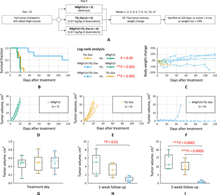
Thermosensitive liposomes represent an important paradigm in oncology, where hyperthermia-mediated release coupled with thermal bioeffects enhance the effectiveness of chemotherapy. Their widespread clinical adoption hinges upon performing controlled targeted hyperthermia, and a leading candidate to achieve this is temperature-based magnetic resonance imaging (MRI)-guided focused ultrasound (MRgFUS). However, the current approach to hyperthermia involves exposures lasting tens of minutes to hours, which is not possible to achieve in many circumstances because of blood vessel cooling and respiratory motion. Here, we investigate a novel approach to overcome these limitations: to use fractionated ultrashort (~30 s) thermal exposures (~41° to 45°C) to release doxorubicin from a thermosensitive liposome. This is first demonstrated in a dorsal chamber tumor model using two-photon microscopy. Thermal exposures were then conducted with a rabbit tumor model using a custom MRgFUS system incorporating temperature feedback control. Drug release was confirmed, and longitudinal experiments demonstrated profoundly enhanced tumor growth inhibition and survival.


Figures

Fig. 1
Fig. 1. Two-photon visualization of doxorubicin release and uptake in mouse tumors during ultrashort FUS thermal exposures.
(A) Illustration of two-photon compatible ring transducer coupled to a FaDu-GFP tumor (black arrows) with implanted thermocouples (white arrows) for temperature feedback. (B) Representative image of the FITC-labeled microcirculation (green) within the FaDu-GFP tumor, whose cells are false-colored magenta under 900-nm excitation. (C) Example temperature profiles within the dorsal window chamber at different target temperatures using real-time feedback control. (D) Changes of the fluorescent signal, indicating the release of doxorubicin, under 810-nm excitation within the FaDu-GFP tumor after heating to hyperthermia temperatures in real time and after 10 ultrashort thermal exposures to 43°C. Tumor cells were not visible under 810-nm excitation. (E) Means ± SD of the intravascular drug signal measured during FUS hyperthermia at each temperature level. Surface plot of the mean doxorubicin fluorescent signal in the extravascular space as a function of distance to the nearest vessel and time, averaged for each mouse tumor heated to (F) 41°, (G) 42°, (H) 43°, and (I) 45°C. (J) Mean fluorescent signal in the extravascular space continues to increase after successive 30 s of heating, indicating that more and more drug is being delivered to the tumor at each hyperthermia temperature elevation. (K) Box-and-whisker plots indicating all data points at the 60-min time point comparing the mean doxorubicin signal in the extravascular compartment at each temperature exposure. The groups were compared using one-way ANOVA with Bonferroni post hoc tests for multiple comparisons; *P < 0.05.
Fig. 2
Fig. 2. MRgFUS treatment planning and tumor temperature profile.
(A) Schematic diagram of the MRgFUS experimental setup. (B) Axial contrast-enhanced T1-weighted MR image of a tumor-bearing rabbit within the MRI-compatible FUS setup. (C) Coronal MR image within the plane of the Vx2 tumor for targeting and MR thermometry feedback purposes. Experimental paradigm, which involves (D) tumor identification, (E) followed by the selection of ROIs within the tumor that will be heated discretely, (F) the administration of TSL-Dox or dextrose alone depending on the treatment group, and (G) real-time MRgFUS heating of the target tumor. Rabbits receiving TSL-Dox alone did not have their tumors heated with MRgFUS. (H) Temperature profile within ROI #4 throughout the entire heat treatment, which consisted of heating all eight ROIs to 42°C for 30 s, 10 times in sequence.
Fig. 3
Fig. 3. MRgFUS temperature control and thermal dose deposition.
Mean temperature ± SD across all Vx2 tumor ROIs used for real-time feedback control (top) and the applied acoustic power to achieve the temperature response (bottom) for the (A) MRgFUS alone group and the (B) MRgFUS+TSL-Dox group in the longitudinal tumor growth study. Tideal was the temperature response that the controller attempted to achieve, and the time values on the horizontal axis are adjusted such that time 0 corresponds to 20 s before FUS was turned on. (C) Example image displaying the thermal dose in CEM43 overlaid upon the anatomical image of the Vx2 tumor that was heated with short-duration MRgFUS hyperthermia. All voxels with a thermal dose above 0.1 CEM43 are displayed in color, and the anatomy is shown in grayscale. (D) Mean temperatures ± SD of the heated ROIs in the heated Vx2 harvested for acute histology. (E) Vx2 tumor identification (white arrows) and ROI selection. (F) Thermal dose following the ultrashort MRgFUS thermal exposures in the heated Vx2 tumor.
Fig. 4
Fig. 4. Doxorubicin distribution in heated and unheated Vx2 tumors.
(A) Experimental timeline for histological examination of heated and unheated Vx2 tumors. DAPI, 4′,6-diamidino-2-phenylindole. (B) Histological examination of bilateral Vx2 tumors, one heated and one unheated under ×10 magnification. Tumors were stained with hematoxylin and eosin (H&E) as well as DAPI for cell nucleus staining, CD31 expression on endothelial cells, and the fluorescence of doxorubicin, all of which were imaged with confocal microscopy. (C) Histological examination of bilateral Vx2 tumors under ×20 magnification. (D) Analysis of the doxorubicin fluorescence as a function of distance to the nearest vessel in heated and unheated tumors. The average data across n = 5 tumor sections spaced 250 μm apart were analyzed for both heated and unheated Vx2 tumors (AU, arbitrary units). (E) The mean fluorescent doxorubicin signal within the Vx2 tumor but outside CD31-stained endothelial cells was compared between all five tumor sections in both heated and unheated Vx2 tumors using an unpaired t test; ***P < 0.001.
Fig. 5
Fig. 5. Vx2 tumor growth following MRgFUS and TSL-Dox.
(A) Survival study design where rabbits were randomly assigned into one of three treatment groups. Following treatment tumor volumes were measured with contrast-enhanced (CE) T1w MR images. (B) Kaplan-Meier survival analysis where groups were compared with a log-rank test and a Bonferroni correction for multiple comparisons; **P < 0.01. (C) Percent change in animal body weight after treatment. Tumor growth curves for the animals receiving MRgFUS alone (D) TSL-Dox alone (E) and the combination of MRgFUS+TSL-Dox (F). Tumor volume comparison on the day of treatment (G) at 1 week after treatment (H) and at 2 weeks following treatment (I). Groups were compared with a one-way ANOVA and a Bonferroni correction for multiple comparisons; *P < 0.05, **P < 0.01, and ***P < 0.001.

References

    1. Yatvin M. B., Weinstein J. N., Dennis W. H., Blumenthal R., Design of liposomes for enhanced local release of drugs by hyperthermia. Science 202, 1290–1293 (1978). - PubMed
    1. Stapleton S., Dunne M., Milosevic M., Tran C. W., Gold M. J., Vedadi A., Mckee T. D., Ohashi P. S., Allen C., Jaffray D. A., Radiation and heat improve the delivery and efficacy of nanotherapeutics by modulating intratumoral fluid dynamics. ACS Nano 12, 7583–7600 (2018). - PubMed
    1. Kong G., Braun R. D., Dewhirst M. W., Hyperthermia enables tumor-specific nanoparticle delivery: Effect of particle size. Cancer Res. 60, 4440–4445 (2000). - PubMed
    1. Sen A., Capitano M. L., Spernyak J. A., Schueckler J. T., Thomas S., Singh A. K., Evans S. S., Hylander B. L., Repasky E. A., Mild elevation of body temperature reduces tumor interstitial fluid pressure and hypoxia and enhances efficacy of radiotherapy in murine tumor models. Cancer Res. 71, 3872–3880 (2011). - PMC - PubMed
    1. Skitzki J. J., Repasky E. A., Evans S. S., Hyperthermia as an immunotherapy strategy for cancer. Curr. Opin. Investig. Drugs 10, 550–558 (2009). - PMC - PubMed

Publication types