. 2020 Jun 17;6(25):eaba2187.
doi: 10.1126/sciadv.aba2187. Print 2020 Jun.

LiQD Cornea: Pro-regeneration collagen mimetics as patches and alternatives to corneal transplantation

Affiliations

LiQD Cornea: Pro-regeneration collagen mimetics as patches and alternatives to corneal transplantation

Christopher D McTiernan et al. Sci Adv. .

Abstract

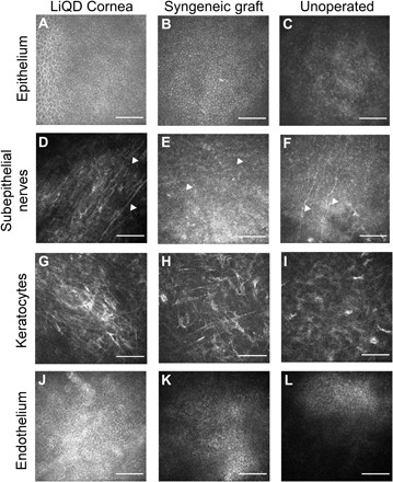
Transplantation with donor corneas is the mainstay for treating corneal blindness, but a severe worldwide shortage necessitates the development of other treatment options. Corneal perforation from infection or inflammation is sealed with cyanoacrylate glue. However, the resulting cytotoxicity requires transplantation. LiQD Cornea is an alternative to conventional corneal transplantation and sealants. It is a cell-free, liquid hydrogel matrix for corneal regeneration, comprising short collagen-like peptides conjugated with polyethylene glycol and mixed with fibrinogen to promote adhesion within tissue defects. Gelation occurs spontaneously at body temperature within 5 min. Light exposure is not required-particularly advantageous because patients with corneal inflammation are typically photophobic. The self-assembling, fully defined, synthetic collagen analog is much less costly than human recombinant collagen and reduces the risk of immune rejection associated with xenogeneic materials. In situ gelation potentially allows for clinical application in outpatient clinics instead of operating theaters, maximizing practicality, and minimizing health care costs.


Figures

Fig. 1
Fig. 1. Biological evaluation of LiQD Cornea.
(A) Immortalized HCECs cultured on LiQD Cornea hydrogels and control tissue culture plastic, showing that the hydrogels support epithelial growth. (B) Expression of T cell costimulatory molecules in BMDCs. Expression of CD40, CD80, and CD86 was measured by flow cytometry, and data are presented as a ratio of mean fluorescence intensity of the experimental samples to untreated BMDCs. LPS acted as a positive control for BMDC activation; *P ≤ 0.05 by Student’s t test. (C) Expression of pro-inflammatory M1 (CD86) and anti-inflammatory M2 (CD206) phenotypic markers at 4 and 7 days after exposure of naïve BMDM precursors to LiQD Cornea hydrogels. (D) Example of a human corneal perforation. (E) Postsurgical photos of rabbits immediately after injecting LiQD Cornea into a perforated cornea. The two-stepped surgically induced perforation can be seen. At day 2 after surgery, the air bubble placed under the cornea during surgery is prominent, indicating that the perforation was completely sealed. The perforated cornea was completed healed by 28 days after operation. Photo credit: Damien Hunter, University of Sydney. (F) Mini-pig corneas where the LiQD Cornea was tested as an alternative to a donor allograft, showing the gross appearance of the LiQD Cornea, syngeneic graft, and an unoperated eye at 12 months after surgery. Photo credit: Monika K. Ljunggren, Linköping University.
Fig. 2
Fig. 2. Clinical exam progression of LiQD Cornea in Göttingen mini-pigs.
(A) Pachymetry showing corneal thickness measured by OCT, showing no significant differences in thickness compared to controls. There was a normal increase in corneal thickness in unoperated controls as the pigs matured. (B) Intraocular pressures were similar in all three groups, showing a slight overall increase over the normal aging process of the pigs. (C) Central corneal haze measured using a modified McDonald-Shadduck scoring system on a scale from 0 to 4. An increase of haze corresponds to the period of in-growth of stromal cells into the cell-free implants. By 12 months after operation, the cells appeared to have attained quiescence. (D) Corneal neovascularization was seen in the LiQD Cornea, mainly from the animal that sustained an unintended perforation. (E) Corneal blink response measured by Cochet-Bonnet esthesiometry showed no significant differences among the three groups. (F) Corneal nerve density in the LiQD Cornea group was significantly lower than the unoperated corneas during months 3 to 9 after operation when the severed nerves were regenerating. (G) Schirmer’s tear test showed similar responses in all three groups tested. (H) Expression of high–molecular weight collagens (HMW, γ, and β), type V collagen, and type I collagen (α1 and α2) in the central portion of the cornea. Figures (A), (B), and (E) to (H) were assessed using a mixed-effects model with a Tukey post hoc test for multiple comparisons. Figures (C) to (D) were analyzed using a Mann-Whitney U test for ordinal data. *P ≤ 0.05 for LiQD Cornea to unoperated, †P ≤ 0.05 for LiQD Cornea to syngeneic graft, and ‡P ≤ 0.05 syngeneic graft to unoperated. All data are plotted as mean ± SEM or mean with individual values.
Fig. 3
Fig. 3. Histopathology, TEM, and immunohistochemistry of the LiQD Cornea at 12 months.
(A to C) Paraffin-embedded sections of porcine cornea stained with hematoxylin and eosin (H&E) show multilayered, nonkeratinizing epithelia in all three samples. (D to F) TEM images of corneal epithelium in all three samples. (G to I) Epithelial cells showed abundance of desmosomes between cells (arrowheads). (J to L) Fully regenerated corneal tear film mucin stained with fluorescein isothiocyanate–conjugated lectin (green) from Ulex europaeus is seen in the LiQD Cornea. This is similar to the tear film in the controls. (M to O) Cytokeratin 12 (red), a marker for fully differentiated corneal epithelial cells, is present in the regenerated LiQD Cornea as in controls. (P to R) CD163 staining (red) shows that a few mononuclear cells are present in stroma of all three samples. Cell nuclei were stained blue with DAPI.
Fig. 4
Fig. 4. EV and exosome secretion of the regenerated LiQD Cornea compared to a healthy unoperated cornea and a syngeneic graft.
(A) Transmission electron micrograph of a LiQD Cornea sample showing the presence of basal epithelial cells invaginations into the stroma. A basement membrane was present. Vesicles can be seen inside the epithelial cell (an example is indicated with a red arrow). EVs are seen (white arrows) in the underlying stromal compartment. (B and C) TEM of syngeneic graft and untreated cornea, respectively. (D to F) Surface reconstructions of corneal sections stained with the cytosolic, EV marker Tsg101 (red) and DAPI (blue). (G to I) Surface reconstruction of colocalized CD9 and Tsg101 staining indicating the presence of exosomes in the basal epithelium and upper stroma of the LiQD Cornea sample. There was less staining in the syngeneic graft and minimal in the untreated control. Scale bars: 500 nm (red) and 20 μm (white).
Fig. 5
Fig. 5. In vivo confocal microscopy images of the LiQD Cornea compared to a healthy unoperated cornea and a syngeneic graft at 12 months after surgery.
Regenerated corneal epithelial cells cover the surface of the LiQD Cornea (A) as with the syngeneic graft (B) and untreated cornea (C). Regenerated nerves (arrowheads) were found at the sub-basal epithelium within the LiQD Cornea (D), ran parallel to one another, and were morphologically similar to those found in the unoperated cornea (F). Nerves in the syngeneic graft were less distinct (E). Keratocytes were present in all corneas (G to I). The unoperated endothelium remained intact and healthy in all corneas (J to L). Scale bars: 100 μm.

References

    1. Oliva M. S., Schottman T., Gulati M., Turning the tide of corneal blindness. Indian J. Ophthalmol. 60, 423–427 (2012). - PMC - PubMed
    1. Gain P., Jullienne R., He Z., Aldossary M., Acquart S., Cognasse F., Thuret G., Global survey of corneal transplantation and eye banking. JAMA Ophthalmol. 134, 167–173 (2016). - PubMed
    1. Jhanji V., Young A. L., Mehta J. S., Sharma N., Agarwal T., Vajpayee R. B., Management of corneal perforation. Surv. Ophthalmol. 56, 522–538 (2011). - PubMed
    1. P. Jarrett, A. Coury, Tissue adhesives and sealants for surgical applications. In Joining and Assembly of Medical Materials and Devices, Y. Zhou, M. D. Breyen, Eds. (Woodhead Publishing, 2013), pp. 449–490.
    1. Vote B. J., Elder M. J., Cyanoacrylate glue for corneal perforations: A description of a surgical technique and a review of the literature. Clin. Exp. Ophthalmol. 28, 437–442 (2000). - PubMed

Publication types

LinkOut - more resources