. 2020 Sep 9;6(37):eaay1422.
doi: 10.1126/sciadv.aay1422. Print 2020 Sep.

3D bioprinting dual-factor releasing and gradient-structured constructs ready to implant for anisotropic cartilage regeneration

Affiliations

3D bioprinting dual-factor releasing and gradient-structured constructs ready to implant for anisotropic cartilage regeneration

Ye Sun et al. Sci Adv. .

Abstract

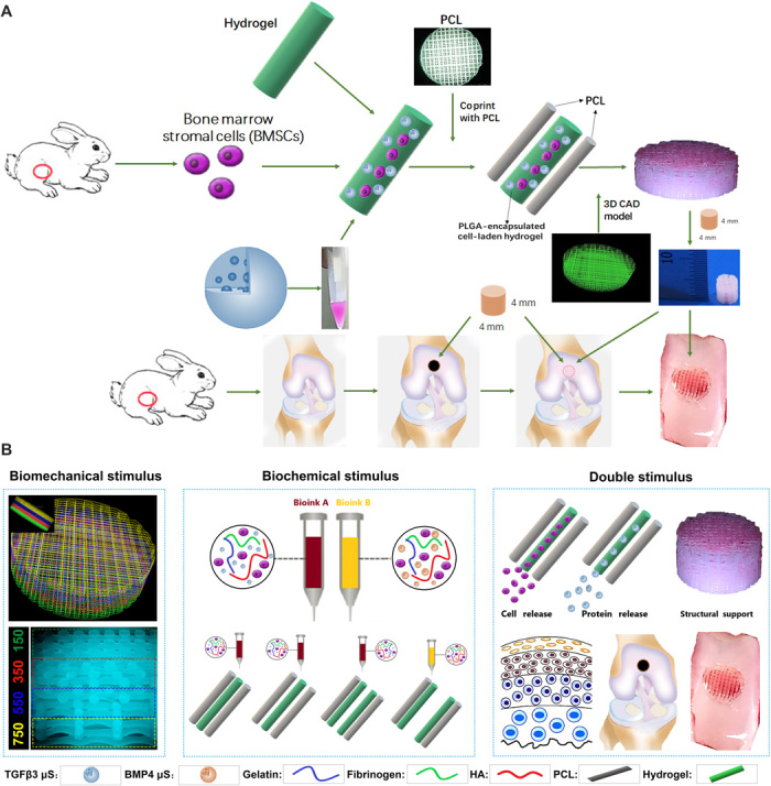
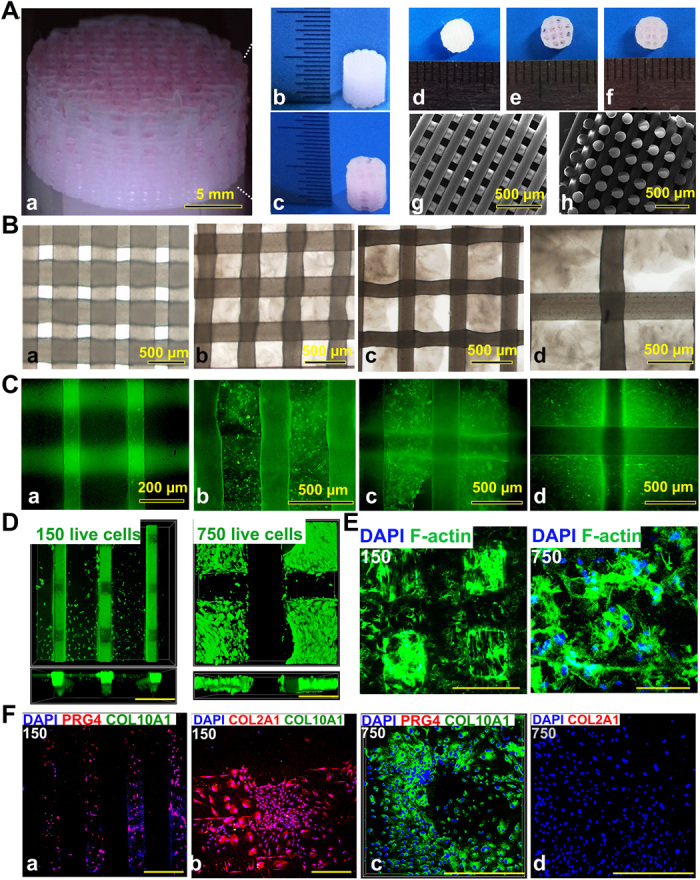
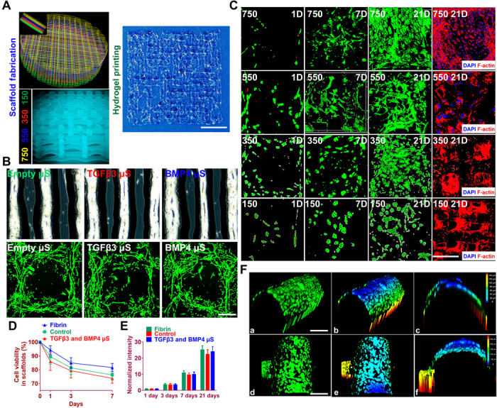
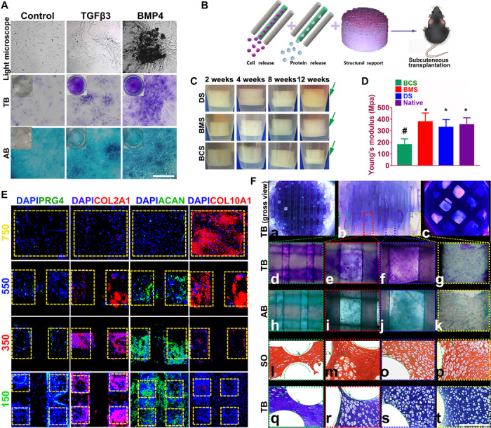
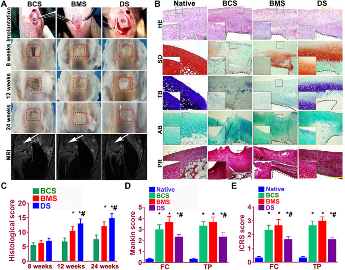
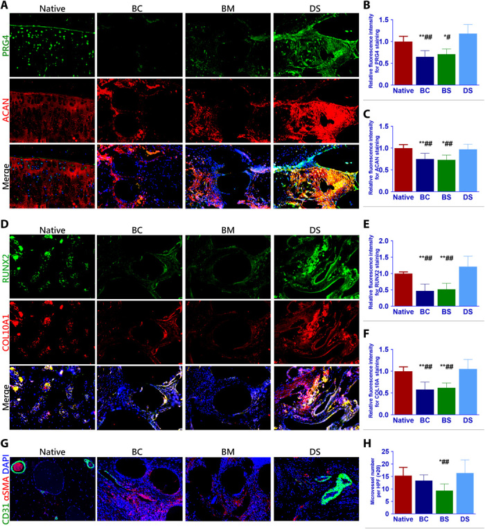
Cartilage injury is extremely common and leads to joint dysfunction. Existing joint prostheses do not remodel with host joint tissue. However, developing large-scale biomimetic anisotropic constructs mimicking native cartilage with structural integrity is challenging. In the present study, we describe anisotropic cartilage regeneration by three-dimensional (3D) bioprinting dual-factor releasing and gradient-structured constructs. Dual-factor releasing mesenchymal stem cell (MSC)-laden hydrogels were used for anisotropic chondrogenic differentiation. Together with physically gradient synthetic biodegradable polymers that impart mechanical strength, the 3D bioprinted anisotropic cartilage constructs demonstrated whole-layer integrity, lubrication of superficial layers, and nutrient supply in deep layers. Evaluation of the cartilage tissue in vitro and in vivo showed tissue maturation and organization that may be sufficient for translation to patients. In conclusion, one-step 3D bioprinted dual-factor releasing and gradient-structured constructs were generated for anisotropic cartilage regeneration, integrating the feasibility of MSC- and 3D bioprinting-based therapy for injured or degenerative joints.


Figures

Fig. 1
Fig. 1. Schematic presentation of the study design and scaffold construction.
(A) Schematic Illustration of the study design with 3D bioprinted dual-factor releasing and gradient-structured MSC-laden constructs for articular cartilage regeneration in rabbits. Schematic diagram of construction of the anisotropic cartilage scaffold and study design. (B) A computer-aided design (CAD) model was used to design the four-layer gradient PCL scaffolding structure to offer BMS for anisotropic chondrogenic differentiation and nutrient supply in deep layers (left). Gradient anisotropic cartilage scaffold was constructed by one-step 3D bioprinting gradient polymeric scaffolding structure and dual protein-releasing composite hydrogels with bioinks encapsulating BMSCs with BMP4 or TGFβ3 μS as BCS for chondrogenesis (middle). The anisotropic cartilage construct provides structural support and sustained release of BMSCs and differentiative proteins for biomimetic regeneration of the anisotropic articular cartilage when transplanted in the animal model (right). Different components in the diagram are depicted at the bottom. HA, hyaluronic acid.
Fig. 2
Fig. 2. 3D bioprinted gradient cartilage scaffold for implantation.
(A) Gross appearance of (a) human-scale and (b and c) rabbit-scale cartilage scaffold (b, NG with 150-μm spacing; c, NG with 750-μm spacing). Top view of the rabbit cartilage scaffold is also shown (d, NG with 150-μm spacing; e, NG with 750-μm spacing; f, gradient scaffold with 150- to 750-μm spacing) atop of the SEM images (g, horizontal section; h, vertical section) taken for the 150-μm NG scaffold to demonstrate the precise alignment of the PCL fibers in the printed scaffold. (B) Deconstruction of the gradient scaffold. The structure of the gradient scaffold was deconstructed into four layers. Microscopic appearance of the hydrogel-PCL composite structure in each layer demonstrated good interconnectivity and delicate, orderly aligned structure for each layer. (C and D) Good cell viability is shown respectively for superficial and deep layers after printing with live/dead assay (green, live cells; red, dead cells) (C) under a microscope and (D) under a confocal microscope. DAPI, 4′,6-diamidino-2-phenylindole. (E) Cell spreading in superficial and deep layers with cytoskeleton staining. (F) Immunostaining for cartilage markers in superficial and deep layers. Expression of COL2A1 and PRG4, the lubrication markers, was significantly higher in the superficial layers with small pore size (a and b), while the chondrogenic cells in the deep layers (c and d) mostly presented with hypertrophic phenotype (COL10A1 expression). Photo credit: Ye Sun, First Affiliated Hospital of Nanjing Medical University.
Fig. 3
Fig. 3. Cell viability and anchoring in the printed anisotropic scaffold.
(A) Schematic of anisotropic cartilage scaffold construction with fabrication of gradient scaffolding structure (left) and large-scale printing of aligned protein-releasing BMSC-laden hydrogel (right). Scale bar, 1 mm. (B) Gross appearance of PLGA μS–encapsulated BMSC-laden hydrogel under a microscope (top). Printed cell-laden hydrogel causes cell alignment in a longitudinal direction of the printed paths, forming a reticular network with cell interaction (bottom). (C) Live/dead cell assays showed ≥95% cell viability maintained through day 1 to 21 for all four layers with gradient spacing (4th row, 150-μm spacing; 3rd row, 350-μm spacing; 2nd row, 550-μm spacing; 1st row, 750-μm spacing). Immunostaining of cytoskeleton (rightmost column) showed cell spreading both in the hydrogel and on the PCL fibers throughout the four layers of the construct. Scale bar, 500 μm. (D and E) Quantified cell viability and proliferation in the printed scaffolds. (F) Cell anchoring in the scaffolds. (a to c) At day 21, good 3D anchoring to the PCL fiber cylinder was observed for the MSC cells released from the hydrogel. (d to f) Similar cell anchoring was observed for PCL fibers in adjacent layers. (b), (c), (e), and (f) are 3D demonstration of cell anchoring in (a) and (d), respectively. Scale bars, 100 μm. Photo credit: Ye Sun, First Affiliated Hospital of Nanjing Medical University.
Fig. 4
Fig. 4. Dual-factor releasing induced cartilaginous matrix formation in 3D bioprinted gradient-structured scaffolds.
(A) Chondrogenic differentiation of condensed rMSCs with toluidine blue (TB) and alcian blue (AB) staining. (B) Scaffolds were transplanted subcutaneously for 12 weeks. (C) To validate the cartilage-generating capability, scaffolds were incubated and observed for 12 weeks in vitro, indicating better cartilage-generating potential for the physically gradient protein-releasing scaffold (movie S2). (D) Young’s modulus of the scaffolds compared with native cartilage after 12 weeks. Data are presented as averages ± SD (n = 6). *P < 0.05 between the NG-750 group and other groups; #P < 0.05 between the native cartilage group and other groups. (E) In the generated cartilage tissues, spatiotemporally released dual-factors induced zone-specific expression of PRG4, aggrecan, and collagens II and X and showed resemblance with native joint cartilage. (F) (a to c) Toluidine blue staining of the 3D printed cartilage constructs (a, top view; b, side view; c, bottom view) after culture in chondrogenic medium for 6 weeks in vitro. (d to g) Toluidine blue and (h to k) alcian blue staining was applied for each layer of the gradient scaffold. (l to p) Safranin O (SO) and (q to t) toluidine blue staining of cartilage tissue between PCL fibers (green curved line) in different layers of the 3D printed cartilage constructs after subcutaneous implantation. Photo credit: Ye Sun, First Affiliated Hospital of Nanjing Medical University.
Fig. 5
Fig. 5. Dual-factor releasing and gradient-structured cartilage scaffold demonstrated better repairing effect of anisotropic cartilage in rabbit knee cartilage defect model in vivo.
(A) Scaffold implantation process and gross appearance of the repair cartilage at 8, 12, and 24 weeks. MRI was made for the operated knee joint (fifth row), demonstrating significant better resolution of subchondral edema and healing of the articular surface (white arrowheads) for joint transplanted with DS scaffolds. (B to F) Chondroprotective effects of the scaffolds were compared by (B) histological scoring evaluation of the repaired cartilage tissue during in vivo implantation. (C) Mankin score and (D) ICRS (International Cartilage Repair Society) histological score of articular cartilage in the femoral condyle (FC) and tibial plateau (TP) in both groups with scaffold implantation. *P < 0.05 between the native group and other groups. #P < 0.05 between the BCS group and the DS group. Data are presented as averages ± SD (N = 6). (A) Histomorphological analysis of the neocartilage tissue at 24 weeks. PR, picrosirius red. The left bottom panels are higher-resolution pictures of the formed neocartilage outline in the colored square boxes. (a to e) Sections were stained with (a) H&E, (b) Safranin O, (c) TB, and (d) AB staining to indicate the presence of proteoglycans in different groups compared with native cartilage. (e) Picrosirius red was used to stain collagens I and III. The brown irregular area at the interface under the formed neocartilage was undegraded PCL material as supporting structure for the scaffolds. Photo credit: Ye Sun, First Affiliated Hospital of Nanjing Medical University.
Fig. 6
Fig. 6. Dual-factor releasing and gradient-structured scaffold restored the anisotropic properties of native cartilage and better microvessel ingrowth.
(A to C) In the superficial layer, immunostaining demonstrated greater PRG4 and ACAN expression in the DS group and the native cartilage compared with other two groups. (D to F) Meanwhile, higher expression of ossification markers (RUNX2 and COL10A1) were also observed for the group with implanted dual-factor releasing and gradient-structured scaffold in deep layers. (G and H) Moreover, the DS scaffold could better promote microvessel ingrowth compared with the group with small pore sizes, indicating better nutrient supply and tissue integration with large pore sizes in the deep zone. *P < 0.05 between the native group and other groups. #P < 0.05 between the DS group and other groups. BC, biochemical stimulus; BS, biomechanical stimulus. **P < 0.01; ##P < 0.01.

References

    1. Freedman B. R., Mooney D. J., Biomaterials to Mimic and Heal Connective Tissues. Adv. Mater. 31, e1806695 (2019). - PMC - PubMed
    1. Lee C. H., Cook J. L., Mendelson A., Moioli E. K., Yao H., Mao J. J., Regeneration of the articular surface of the rabbit synovial joint by cell homing: a proof of concept study. Lancet 376, 440–448 (2010). - PMC - PubMed
    1. Merriam A. R., Patel J. M., Culp B. M., Gatt C. J. Jr., Dunn M. G., Successful Total Meniscus Reconstruction Using a Novel Fiber-Reinforced Scaffold: A 16- and 32-Week Study in an Ovine Model. Am. J. Sports Med. 43, 2528–2537 (2015). - PubMed
    1. Ayers D. C., How Common Is Revision for Adverse Reaction to Metal Debris After Total Hip Replacement with a Metal-on-Polyethylene Bearing Surface?: Commentary on an article by Anders Persson, MD, et al.: "Revision for Symptomatic Pseudotumor Following Primary Total Hip Arthroplasty with a Standard Femoral Stem". J. Bone Joint Surg. Am. 100, e82 (2018). - PubMed
    1. Craft A. M., Rockel J. S., Nartiss Y., Kandel R. A., Alman B. A., Keller G. M., Generation of articular chondrocytes from human pluripotent stem cells. Nat. Biotechnol. 33, 638–645 (2015). - PubMed

Publication types

LinkOut - more resources