. 2020 Sep 11;6(37):eabb1611.
doi: 10.1126/sciadv.abb1611. Print 2020 Sep.

Dimorphism in cryptophytes-The case of Teleaulax amphioxeia/ Plagioselmis prolonga and its ecological implications

Affiliations

Dimorphism in cryptophytes-The case of Teleaulax amphioxeia/ Plagioselmis prolonga and its ecological implications

A Altenburger et al. Sci Adv. .

Abstract

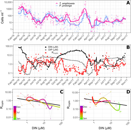
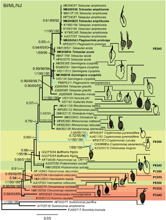
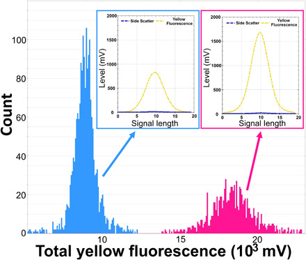
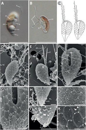
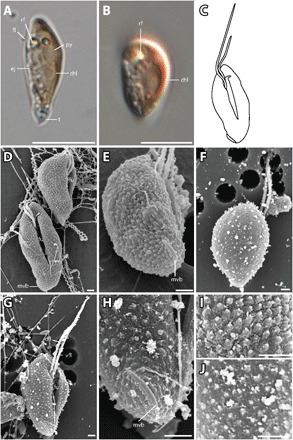
Growing evidence suggests that sexual reproduction might be common in unicellular organisms, but observations are sparse. Limited knowledge of sexual reproduction constrains understanding of protist ecology. Although Teleaulax amphioxeia and Plagioselmis prolonga are common marine cryptophytes worldwide, and are also important plastid donors for some kleptoplastic ciliates and dinoflagellates, the ecology and development of these protists are poorly known. We demonstrate that P. prolonga is the haploid form of the diploid T. amphioxeia and describe the seasonal dynamics of these two life stages. The diploid T. amphioxeia dominates during periods of high dissolved inorganic nitrogen (DIN) and low irradiance, temperature, and grazing (winter and early spring), whereas the haploid P. prolonga becomes more abundant during the summer, when DIN is low and irradiance, temperature, and grazing are high. Dimorphic sexual life cycles might explain the success of this species by fostering high genetic diversity and enabling endurance in adverse conditions.


Figures

Fig. 1
Fig. 1. Dynamics of Plagioselmis prolonga and Teleaulax amphioxeia.
(A) Dynamics of cell abundance of P. prolonga (blue circles) and T. amphioxeia (pink triangles) in Roskilde Fjord from March 2016 to November 2017. (B) Dynamics of Rcrypto, the ratio between P. prolonga to T. amphioxeia (red triangles), DIN (black circles), and DIP (gray circles) concentration for the same period depicted in (A). (C and D) Trajectory dynamics between Rcrypto and DIN concentrations in 2016 (C) and 2017 (D). In (A) and (B), symbols are the observations, and lines are the 10 days moving average. In (C) and (D), dots represent the observations, colored thick lines represent the time trajectories estimated from the GAM, and the thick black lines represent the linear relationship between DIN and Rcrypto. Trajectories for the 2 years are shown separately for better visualizing the idiosyncrasy.
Fig. 2
Fig. 2. Phylogeny of cryptophytes analyzed by Bayesian inference of 18S rDNA sequences.
Numbers at nodes represent the posterior probability of Bayesian analysis/bootstrap values of maximum likelihood out of 1000 replicates/bootstrap values of neighbor-joining out of 10,000 replicates. Not resolved branches and branches with support values below 50% are marked with a minus sign (−). The scale bar corresponds to three substitutions per 100 nucleotide positions. Colors correspond to absorption maxima (i.e., PE545 = green). Branches for freshwater species are blue, marine and brackish species are black. Newly sequenced strains are in bold. Drawings of cells are not to scale.
Fig. 3
Fig. 3. Nuclei fluorescence of P. prolonga and T. amphioxeia.
Histograms showing the nuclei counts in different classes of total yellow fluorescence (FLY) (proxy for amount of DNA) in extracted nuclei of P. prolonga (blue) and T. amphioxeia (pink) cultures stained with SYBR green dye. An example of an optical profile for the nuclei of each species is also provided, showing the high FLY and low SWS signal (characteristic for the nuclei) and differences in the FLY intensity between the two forms.
Fig. 4
Fig. 4. P. prolonga morphology.
(A) Lateral view of P. prolonga with the tail (t), nucleus (nu), ejectosomes (ej), pyrenoid (py), and flagellum (fl). (B) Lateral view of P. prolonga without the tail, with chloroplast (chl), ejectosomes, and flagella. (C) Drawings of P. prolonga cell with the tail (left) and without the tail (right). (D) Lateral view with part of the surface periplast component (SPC). (E) Lateral view showing the inner periplast component (IPC) and tail; note the hexagonal periplast plates of the IPC and the ventral flagellum (vfl), which is shorter than the dorsal flagellum (dfl). (F) Ventral view of a cell without tail. The furrow (fu), IPC, flagella, and the mid-ventral band (mvb) are visible. (G) Closeup of (C) with detail of the mid-ventral band that bifurcates at the base of the furrow. (H) The mid-ventral band terminates before reaching the furrow and does not bifurcate. (I) Detail of the posterior end of a cell without tail. There are no hexagonal plates at the posterior end, and the mid-ventral band extends onto the dorsal side of the cell (arrow). (J) Detailed view of the periplast; the particles are probably part of the SPC. (K) Detailed view of the IPC. Scale bars are 10 μm (light microscopy) and 1 μm [scanning electron microscopy (SEM)]. Anterior of all cell oriented upwards.
Fig. 5
Fig. 5. T. amphioxeia morphology.
(A) Lateral view of elongated T. amphioxeia cell with olive-brown color with chloroplast, refractosome (rf), pyrenoid, and ejectosomes. (B) Lateral view of a more round T. amphioxeia cell, more reddish in color with chloroplast and refractosome. (C) Drawing of T. amphioxeia surface characters. (D) Ventral and lateral view of cells with intact SPC with a papillate structure (fixation with Lugol’s solution), furrow, and mid-ventral band that is almost lateral at the antapical end. (E) View from lateroposterior on the cell with SPC showing the short mid-ventral band. (F) Dorsal view of the cell with sheet-like IPC (fixation with OsO4). (G) Ventral view showing the furrow and ventral flagellum that is longer than the dorsal flagellum. (H) Detail of the antapical end with the mid-ventral band, which is short and curved and does not extend to the furrow. (I) Detail of the cell surface SPC visible in fixation with Lugol’s solution. (J) Detail of the cell surface IPC in fixation with OsO4. Scale bars are 10 μm (light microscopy) and 1 μm (SEM). Anterior of all cell oriented upwards.

References

    1. Cerino F., Zingone A., A survey of cryptomonad diversity and seasonality at a coastal Mediterranean site. Eur. J. Phycol. 41, 363–378 (2006).
    1. Gustafson D. E., Stoecker D. K., Johnson M. D., Van Heukelem W. F., Sneider K., Cryptophyte algae are robbed of their organelles by the marine ciliate Mesodinium rubrum. Nature 405, 1049–1052 (2000). - PubMed
    1. Hansen P. J., Moldrup M., Tarangkoon W., Garcia-Cuetos L., Moestrup Ø., Direct evidence for symbiont sequestration in the marine red tide ciliate Mesodinium rubrum. Aquat. Microb. Ecol. 66, 63–75 (2012).
    1. Koike K., Takishita K., Anucleated cryptophyte vestiges in the gonyaulacalean dinoflagellates Amylax buxus and Amylax triacantha (Dinophyceae). Phycol. Res. 56, 301–311 (2008).
    1. Park M. G., Kim M., Kang M., A dinoflagellate Amylax triacantha with plastids of the cryptophyte origin: phylogeny, feeding mechanism, and growth and grazing responses. J. Eukaryot. Microbiol. 60, 363–376 (2013). - PubMed

Publication types

LinkOut - more resources