. 2020 Sep 11;6(37):eabb2630.
doi: 10.1126/sciadv.abb2630. Print 2020 Sep.

Extracellular matrix stiffness determines DNA repair efficiency and cellular sensitivity to genotoxic agents

Affiliations

Extracellular matrix stiffness determines DNA repair efficiency and cellular sensitivity to genotoxic agents

Min Deng et al. Sci Adv. .

Abstract

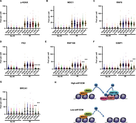
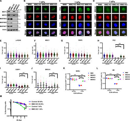
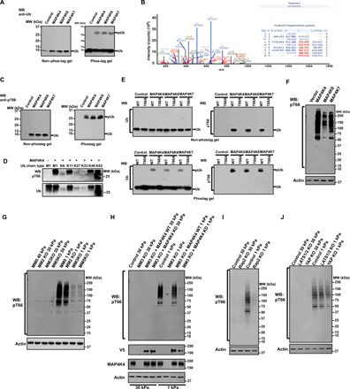
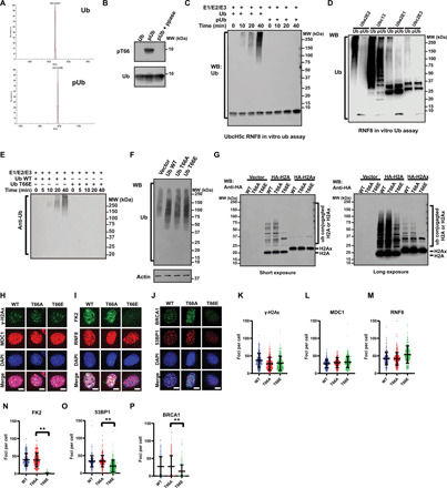
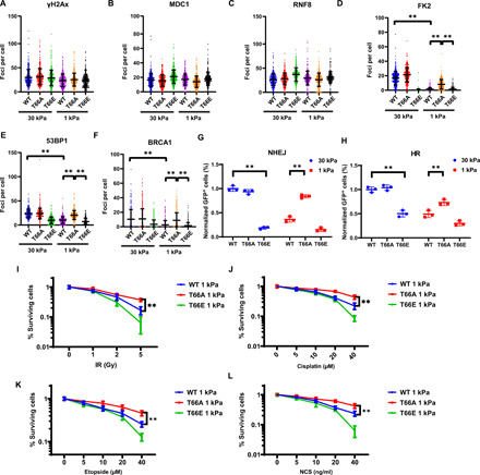
DNA double-strand breaks (DSBs) are highly toxic lesions that can drive genetic instability. These lesions also contribute to the efficacy of radiotherapy and many cancer chemotherapeutics. DNA repair efficiency is regulated by both intracellular and extracellular chemical signals. However, it is largely unknown whether this process is regulated by physical stimuli such as extracellular mechanical signals. Here, we report that DSB repair is regulated by extracellular mechanical signals. Low extracellular matrix (ECM) stiffness impairs DSB repair and renders cells sensitive to genotoxic agents. Mechanistically, we found that the MAP4K4/6/7 kinases are activated and phosphorylate ubiquitin in cells at low stiffness. Phosphorylated ubiquitin impairs RNF8-mediated ubiquitin signaling at DSB sites, leading to DSB repair deficiency. Our results thus demonstrate that ECM stiffness regulates DSB repair efficiency and genotoxic sensitivity through MAP4K4/6/7 kinase-mediated ubiquitin phosphorylation, providing a previously unidentified regulation in DSB-induced ubiquitin signaling.


Figures

Fig. 1
Fig. 1. Low stiffness impairs DSB repair and increases cellular sensitivity to genotoxic agents.
(A) HEK293 cells grown on different surfaces were stained for F-actin and nucleus. DAPI, 4′,6-diamidino-2-phenylindole. (B) The elasticity of hydrogels was measured by atomic force microscopy. (C) The coating efficiency of fibronectin on hydrogels was measured. (D to H) HEK293 cells grown on different surfaces were treated with indicated doses of genotoxic agents or Palbociclib. Colony formation assays were performed to detect cell survival. (I) HEK293 cells grown on different surfaces were treated with indicated genotoxic agents [10 Gy IR, 10 μM cisplatin, 10 μM etoposide, and NCS (10 ng/ml)]. Cells were trypsinized after 48 hours and cellular apoptosis was detected by annexin V and PI staining. (J) HEK293 cells grown on different surfaces were treated with IR (2 Gy) and stained for γ-H2AX at the indicated time points. (K) HEK293 cells grown on different surfaces were treated with IR (10 Gy) and harvested at the indicated time points for neutral comet assay. Data are presented as means ± SEM. More than 50 cells per group were quantified (**P < 0.01). (L and M) HEK293 cells with chromosomally integrated HR or NHEJ reporter were plated on different surfaces. The efficiency of NHEJ (L) and HR (M) were analyzed by flow cytometry.
Fig. 2
Fig. 2. Low stiffness inhibits DSB repair at the level of RNF8 in the DSB repair pathway.
(A to G) HEK293 cells were grown for 24 hours on fibronectin-coated hydrogels of different stiffness. Cells were fixed 1 hour after irradiation (1 Gy) and stained with anti–γ-H2AX (A), MDC1 (B), RNF8 (C), FK2 (D), 53BP1 (F), and BRCA1(G) antibodies. For RNF168 (E), HEK293 cells expressing mCherry-RNF168 were plated on fibronectin-coated hydrogels of different stiffness. Cells were fixed 1 hour after irradiation (1 Gy), and RNF168 foci were visualized with mCherry. Quantification is described in Methods. Data are presented as means ± SD, n = 3 biologically independent samples (**P < 0.01). (H) A model showing the affected DNA repair steps by low stiffness.
Fig. 3
Fig. 3. Inhibition of DSB repair by low stiffness is dependent on MAP4K4/6/7.
(A) Western blots showing the expression levels of MST1, MST2, MAP4K4, MAP4K6, and MAP4K7 in control (MM0), MM2KO, MM3KO, and MM5KO HEK293 cells. Mw, weight average molecular weight. (B to D) MM0, MM2KO, MM3KO, and MM5KO cells were grown on soft (1 kPa) and stiff (30 kPa) fibronectin-coated hydrogels. Cells were fixed 1 hour after irradiation (1 Gy) and stained with anti–γ-H2AX and MDC1 (B), RNF8 and FK2 (C), and 53BP1 and BRCA1 (D) antibodies. Scale bars, 10 μM. (E to H) Quantification of (B) to (D) is described in Methods. (K and L) MM0, MM2KO, MM3KO, and MM5KO cells were grown on soft (1 kPa) and stiff (30 kPa) fibronectin-coated hydrogels. Effect of ECM stiffness on the efficiency of NHEJ (K) and HR (L) in indicated cells was analyzed by flow cytometry. (M) MAP4K4/6/7 kinases are required for regulation of stiffness-induced radiation sensitivity. Colony formation assays were performed to examine survival of WT (MM0) and MM3KO HEK293 cells on soft (1 kPa) and stiff (30 kPa) fibronectin-coated hydrogels when exposed to the indicated doses of radiation.
Fig. 4
Fig. 4. MAP4K4/6/7 kinases phosphorylate ubiquitin in vitro and in cells.
(A) An in vitro kinase assay was performed at 30°C for 1 hour in the presence of free Ub and indicated purified kinases. Samples were run on non–phos-tag or phos-tag PAGE and the gels were blotted with anti-Ub antibody. WB, Western blotting; MW, molecular weight. (B) Gel band of phosphorylated ubiquitin was trypsinized and subjected to LC-MS/MS analysis. (C) Samples were processed as in (A) and blotted with anti-pT66 antibody. (D) An in vitro kinase assay was performed with different tetra-Ub chains and MAP4K4. The gels were subjected to Western blot with anti-pT66 antibody (top) or anti-Ub antibody (bottom). (E) An in vitro kinase assay was performed with indicated Ub proteins and purified kinases. Samples were run on non–phos-tag or phos-tag PAGE, and the gels were blotted with anti-Ub or anti-pT66 antibody. WT, wild type. (F) HEK293 cells grown on plastic plates were transfected with the indicated constructs, and phosphorylation of ubiquitin in cells was detected with anti-pT66 antibody. Actin was used as a loading control. (G to J) Indicated cells were cultured on low (1 kPa) and high (30 kPa) stiffness fibronectin-coated hydrogels for 24 hours. Cell lysates were probed with indicated antibodies. Actin was used as loading control.
Fig. 5
Fig. 5. Phosphorylation of ubiquitin blocks RNF8/RNF168-mediated ubiquitin conjugation in vitro and in cells.
(A) Purified Ub variants were analyzed by electrospray ionization mass spectrometry. (B) Ub variants were treated with or without λ-PPase and blotted with indicated antibodies. (C) The assembly of ubiquitin chains was determined in the presence of Ube1, UbcH5c, and RNF8 and indicated Ub variants. Samples were taken at the indicated time and immunoblotted with anti-Ub antibody. (D) The assembly of ubiquitin chains was determined in the presence of Ube1, indicated E2s, RNF8, and indicated Ub variants. Polyubiquitin chains were detected by immunoblotting with an anti-Ub antibody.(E) The assembly of ubiquitin chains was determined in the presence of Ube1, UbcH5c, RNF8, and indicated Ub variants. Samples were taken at the indicated time and immunoblotted with anti-Ub antibody. (F) HEK293 expressing WT, T66A, and T66E mutant ubiquitin were blotted with anti-Ub antibody. (G) Cells as in (F) were transfected with indicated plasmids. Cells were irradiated (10 Gy) and blotted with anti-HA antibody. (H to J) Cells as in (F) were grown on glass cover slips. Cells were fixed 1 hour after irradiation (1 Gy) and stained with indicated antibodies. (K to P) Quantification of (H) to (J). **P < 0.01.
Fig. 6
Fig. 6. Phosphorylation of ubiquitin mediated DNA repair blockage in cells at low stiffness.
(A to F) Ubiquitin-replacement HEK293 cells expressing wild-type (WT), T66A, or T66E mutant ubiquitin were plated on soft (1 kPa) and stiff (30 kPa) fibronectin-coated hydrogels. Cells were fixed 1 hour after irradiation and stained with anti–γ-H2AX (A), MDC1 (B), RNF8 (C), FK2 (D), 53BP1 (E), and BRCA1 (F) antibodies. Data are presented as means ± SD, n = 3 biologically independent samples (**P < 0.01). (G and H) Phosphorylation of ubiquitin inhibits HR and NHEJ. Effect of ubiquitin phosphorylation on the efficiency of NHEJ (G) and HR (H) was analyzed by flow cytometry. Data are presented as means ± SD. n = 3 biologically independent samples (**P < 0.01). (I to L) Phosphorylation of ubiquitin regulates genotoxic sensitivity. Ubiquitin-replacement HEK293 cells expressing wild-type (WT), T66A, or T66E mutant ubiquitin were plated on soft (1 kPa) fibronectin-coated hydrogels. Cells were treated with indicated genotoxic agents. Colony formation assays were performed to examine survival of cells expressing wild-type (WT), T66A, or T66E mutant ubiquitin on soft (1 kPa) fibronectin-coated hydrogels. Data are presented as means ± SD. n = 3 biologically independent samples.

References

    1. Reinhardt H. C., Yaffe M. B., Phospho-Ser/Thr-binding domains: Navigating the cell cycle and DNA damage response. Nat. Rev. Mol. Cell Biol. 14, 563–580 (2013). - PubMed
    1. Bassing C. H., Chua K. F., Sekiguchi J., Suh H., Whitlow S. R., Fleming J. C., Monroe B. C., Ciccone D. N., Yan C., Vlasakova K., Livingston D. M., Ferguson D. O., Scully R., Alt F. W., Increased ionizing radiation sensitivity and genomic instability in the absence of histone H2AX. Proc. Natl. Acad. Sci. U.S.A. 99, 8173–8178 (2002). - PMC - PubMed
    1. Celeste A., Petersen S., Romanienko P. J., Fernandez-Capetillo O., Chen H. T., Sedelnikova O. A., Reina-San-Martin B., Coppola V., Meffre E., Difilippantonio M. J., Redon C., Pilch D. R., Olaru A., Eckhaus M., Camerini-Otero R. D., Tessarollo L., Livak F., Manova K., Bonner W. M., Nussenzweig M. C., Nussenzweig A., Genomic instability in mice lacking histone H2AX. Science 296, 922–927 (2002). - PMC - PubMed
    1. Rogakou E. P., Pilch D. R., Orr A. H., Ivanova V. S., Bonner W. M., DNA double-stranded breaks induce histone H2AX phosphorylation on serine 139. J. Biol. Chem. 273, 5858–5868 (1998). - PubMed
    1. Stewart G. S., Wang B., Bignell C. R., Taylor A. M. R., Elledge S. J., MDC1 is a mediator of the mammalian DNA damage checkpoint. Nature 421, 961–966 (2003). - PubMed

Publication types

LinkOut - more resources