. 2020 Sep 11;92(1):45-52.
doi: 10.1136/jnnp-2020-323894. Online ahead of print.

In vivo neuroinflammation and cerebral small vessel disease in mild cognitive impairment and Alzheimer's disease

Affiliations

In vivo neuroinflammation and cerebral small vessel disease in mild cognitive impairment and Alzheimer's disease

Audrey Low et al. J Neurol Neurosurg Psychiatry. .

Abstract

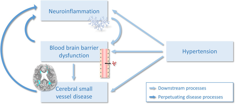
Introduction: Associations between cerebral small vessel disease (SVD) and inflammation have been largely examined using peripheral blood markers of inflammation, with few studies measuring inflammation within the brain. We investigated the cross-sectional relationship between SVD and in vivo neuroinflammation using [11C]PK11195 positron emission tomography (PET) imaging.

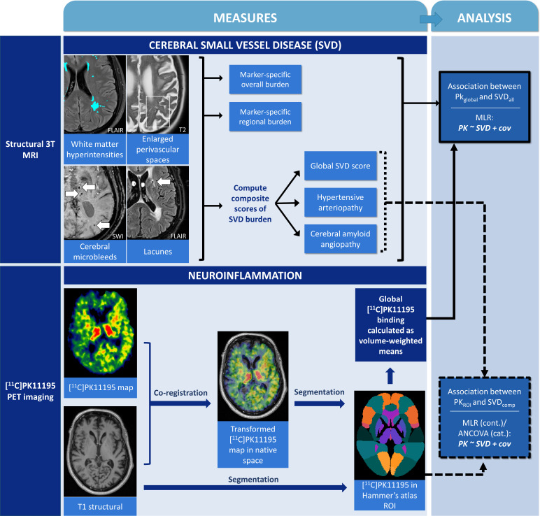
Methods: Forty-two participants were recruited (according to NIA-AA guidelines, 14 healthy controls, 14 mild Alzheimer's disease, 14 amyloid-positive mild cognitive impairment). Neuroinflammation was assessed using [11C]PK11195 PET imaging, a marker of microglial activation. To quantify SVD, we assessed white matter hyperintensities (WMH), enlarged perivascular spaces, cerebral microbleeds and lacunes. Composite scores were calculated for global SVD burden, and SVD subtypes of hypertensive arteriopathy and cerebral amyloid angiopathy (CAA). General linear models examined associations between SVD and [11C]PK11195, adjusting for sex, age, education, cognition, scan interval, and corrected for multiple comparisons via false discovery rate (FDR). Dominance analysis directly compared the relative importance of hypertensive arteriopathy and CAA scores as predictors of [11C]PK11195.

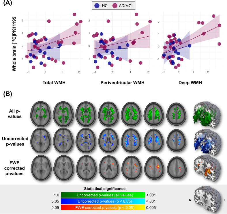
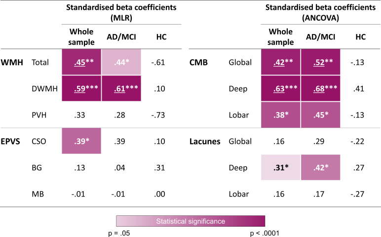
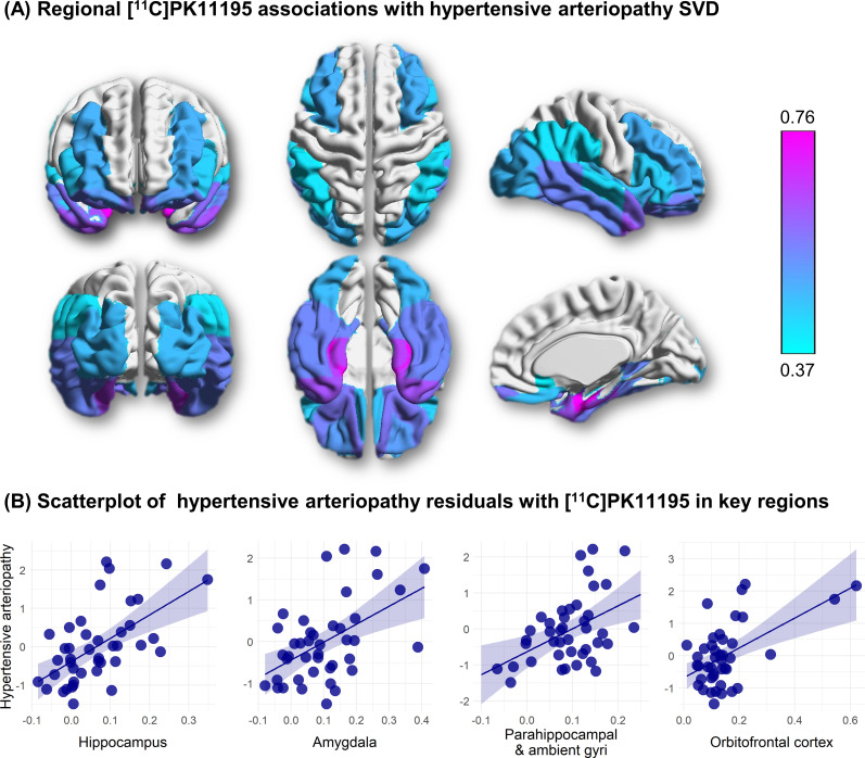
Results: Global [11C]PK11195 binding was associated with SVD markers, particularly in regions typical of hypertensive arteriopathy: deep microbleeds (β=0.63, F(1,35)=35.24, p<0.001), deep WMH (β=0.59, t=4.91, p<0.001). In dominance analysis, hypertensive arteriopathy score outperformed CAA in predicting [11C]PK11195 binding globally and in 28 out of 37 regions of interest, especially the medial temporal lobe (β=0.66-0.76, t=3.90-5.58, FDR-corrected p (pFDR)=<0.001-0.002) and orbitofrontal cortex (β=0.51-0.57, t=3.53-4.30, pFDR=0.001-0.004).

Conclusion: Microglial activation is associated with SVD, particularly with the hypertensive arteriopathy subtype of SVD. Although further research is needed to determine causality, our study suggests that targeting neuroinflammation might represent a novel therapeutic strategy for SVD.


Conflict of interest statement

Competing interests: None declared.

Figures

Figure 1
Figure 1
Schematic representation of multimodal imaging data, image processing pipeline, quantification of pathological burden and statistical approach to test associations between pathologies. ACE-R, Addenbrooke’s Cognitive Examination-Revised; cat., categorical measures, for example, presence/absence of microbleeds and lacunes; cov, covariates (sex, age, education, ACE-R, and scan interval); cont., continuous SVD measures; MLR, multiple linear regression; PKglobal, global [11C]PK11195 binding; PKROI, [11C]PK11195 binding in each Hammer’s atlas region of interest; SVDall, each measure of SVD, including global and regional data of each SVD marker, and composite SVD scores; SVDcomp, composite SVD scores; SVD, small vessel disease.
Figure 2
Figure 2
Association between whole brain [11C]PK11195 binding and WMH. (A) Scatterplot of whole brain [11C]PK11195 binding with WMH volumes. WMH values are residuals adjusted for sex, age, education, ACE-R score and scan interval. (B) Lesion probability maps of [11C]PK11195-related WMH spatial distribution, adjusted for sex, age, education, ACE-R score and scan interval. ACE-R, Addenbrooke’s Cognitive Examination-Revised; AD, Alzheimer’s disease; FWE, family-wise error; HC, healthy controls; MCI, mild cognitive impairment; WMH, white matter hyperintensities.
Figure 3
Figure 3
Graphical summary of associations between global [11C]PK11195 and SVD across imaging markers and regions. Values represent standardised beta coefficients of the individual markers, adjusted for sex, age, education, ACE-R and scan interval. Multiple linear regression was conducted for WMH and EPVS, while ANCOVA was conducted for presence/absence of CMB and lacunes. Colour scale represents p values, whereby darker shades signify smaller p values and unshaded (white) cells are not statistically significant. *p<0.05, **p<0.01, ***p<0.001; bold values represent statistical significance (p<0.05) before correcting for multiple comparisons; underlined values represent statistical significance (p<0.05) after false discovery rate correction for multiple comparisons (12 measures × 3 groups=36 comparisons). ACE-R, Addenbrooke’s Cognitive Examination-Revised; ANCOVA, analysis of covariance; AD/MCI, participants with Alzheimer’s disease or mild cognitive impairment; BG, basal ganglia; CMB, cerebral microbleeds; CSO, centrum semiovale; DWMH, deep white matter hyperintensities; HC, healthy controls; EPVS, enlarged perivascular spaces; MLR, multiple linear regression; PVH, periventricular white matter hyperintensities; MB, midbrain; SVD, small vessel disease; WMH, white matter hyperintensities.
Figure 4
Figure 4
Associations between regional [11C]PK11195 binding and hypertensive arteriopathy (n=42). (A) β-weight brain mapping of associations between regional [11C]PK11195 binding and hypertensive arteriopathy. Coloured overlay of the Hammers atlas represents statistically significant elevation of [11C]PK11195 binding with hypertensive arteriopathy score controlling for sex, age, education, ACE-R score and scan interval and corrected for multiple comparisons. Colour gradient represents the strength of association (standardised β weights), increasing in magnitude from light blue to fuchsia. (B) Scatterplots of relationships between hypertensive arteriopathy (y-axis) and [11C]PK11195 binding in key regions of interest (x-axis). Hypertensive arteriopathy values are residuals, adjusted for sex, age, education, ACE-R and scan interval. ACE-R, Addenbrooke’s Cognitive Examination-Revised; SVD, small vessel disease.
Figure 5
Figure 5
The vicious cycle of neuroinflammation and cerebral small vessel disease.

References

    1. Bos D, Wolters FJ, Darweesh SKL, et al. . Cerebral small vessel disease and the risk of dementia: a systematic review and meta-analysis of population-based evidence. Alzheimers Dement 2018;14:1482–92. 10.1016/j.jalz.2018.04.007 - DOI - PubMed
    1. Heneka MT, Carson MJ, El Khoury J, et al. . Neuroinflammation in Alzheimer's disease. Lancet Neurol 2015;14:388–405. 10.1016/S1474-4422(15)70016-5 - DOI - PMC - PubMed
    1. Kinney JW, Bemiller SM, Murtishaw AS, et al. . Inflammation as a central mechanism in Alzheimer's disease. Alzheimers Dement 2018;4:575–90. 10.1016/j.trci.2018.06.014 - DOI - PMC - PubMed
    1. Passamonti L, Rodríguez PV, Hong YT, et al. . [11C]PK11195 binding in Alzheimer disease and progressive supranuclear palsy. Neurology 2018;90:e1989–96. 10.1212/WNL.0000000000005610 - DOI - PMC - PubMed
    1. Stefaniak J, O'Brien J. Imaging of neuroinflammation in dementia: a review. J Neurol Neurosurg Psychiatry 2016;87:21–8. 10.1136/jnnp-2015-311336 - DOI - PubMed

LinkOut - more resources