. 2020 Sep 11;11(1):4571.
doi: 10.1038/s41467-020-18405-9.

Pre-symptomatic Caspase-1 inhibitor delays cognitive decline in a mouse model of Alzheimer disease and aging

Affiliations

Pre-symptomatic Caspase-1 inhibitor delays cognitive decline in a mouse model of Alzheimer disease and aging

Joseph Flores et al. Nat Commun. .

Erratum in

Abstract

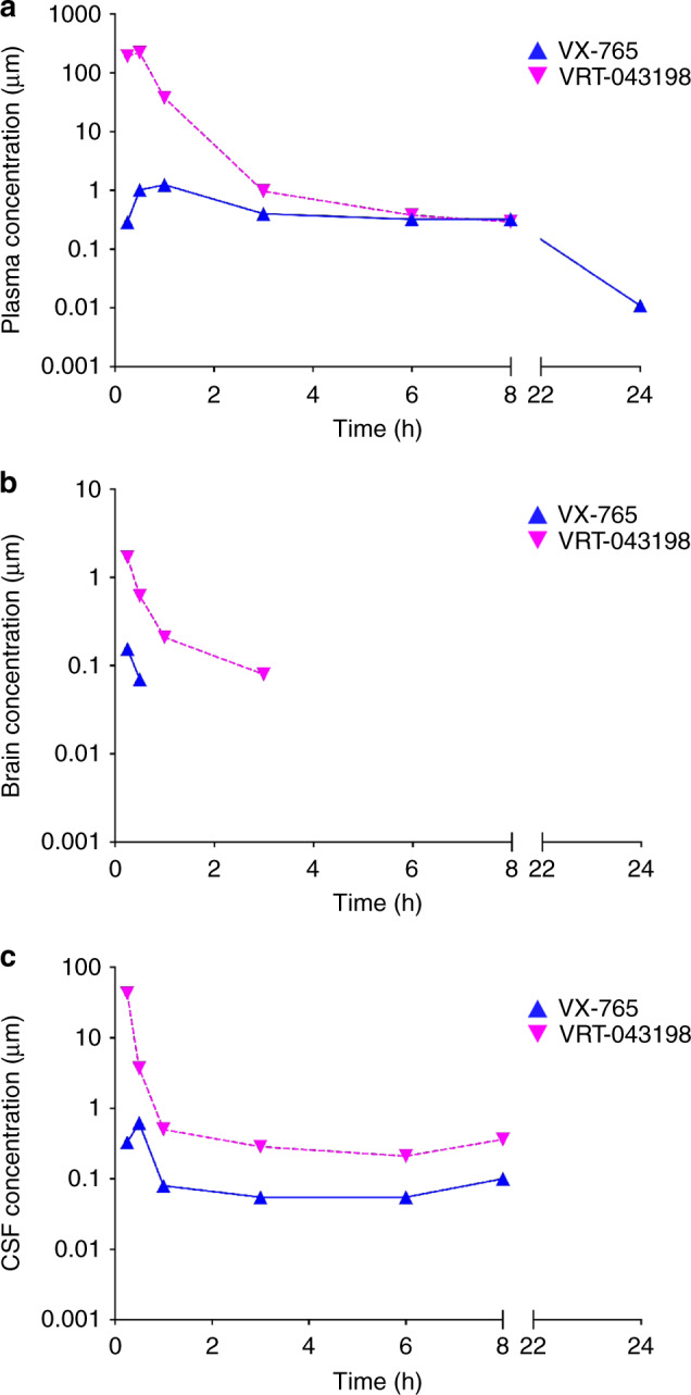
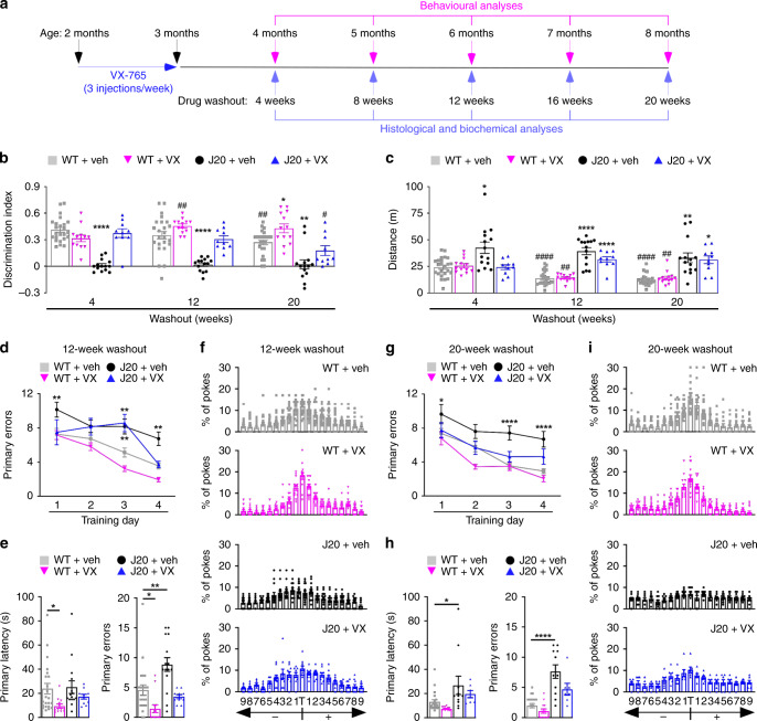
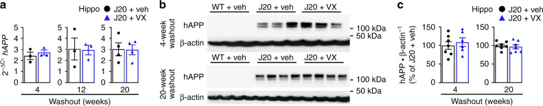
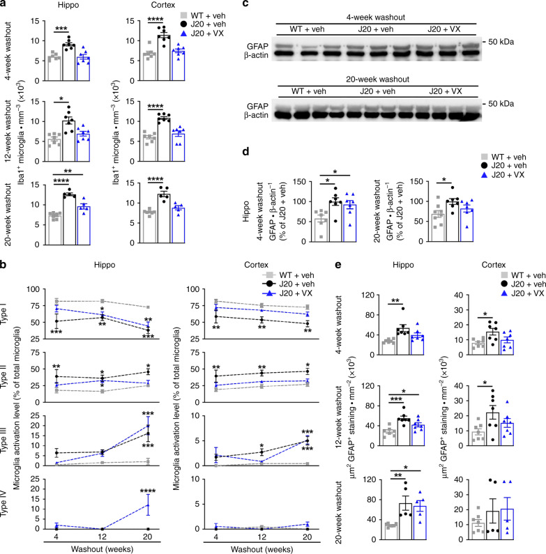
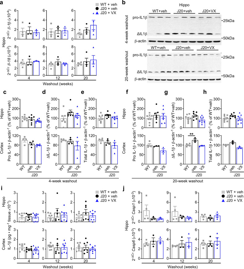
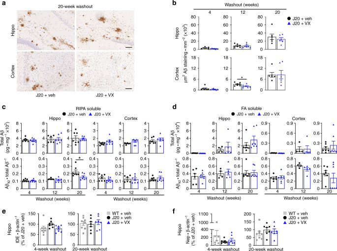
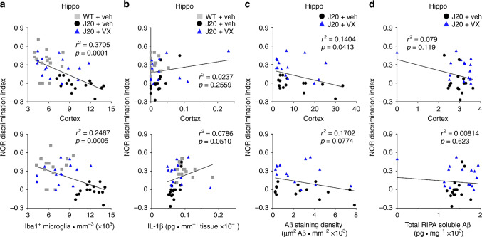
Early therapeutic interventions are essential to prevent Alzheimer Disease (AD). The association of several inflammation-related genetic markers with AD and the early activation of pro-inflammatory pathways in AD suggest inflammation as a plausible therapeutic target. Inflammatory Caspase-1 has a significant impact on AD-like pathophysiology and Caspase-1 inhibitor, VX-765, reverses cognitive deficits in AD mouse models. Here, a one-month pre-symptomatic treatment of Swedish/Indiana mutant amyloid precursor protein (APPSw/Ind) J20 and wild-type mice with VX-765 delays both APPSw/Ind- and age-induced episodic and spatial memory deficits. VX-765 delays inflammation without considerably affecting soluble and aggregated amyloid beta peptide (Aβ) levels. Episodic memory scores correlate negatively with microglial activation. These results suggest that Caspase-1-mediated inflammation occurs early in the disease and raise hope that VX-765, a previously Food and Drug Administration-approved drug for human CNS clinical trials, may be a useful drug to prevent the onset of cognitive deficits and brain inflammation in AD.


Conflict of interest statement

The authors declare no competing interests.

Figures

Fig. 1
Fig. 1. VX-765 and VRT-043198 are detected in mice after a single dose of VX-765.
ac PK measures of VX-765 and active VRT-043198 metabolite in a plasma, b whole brain homogenates, and c CSF samples up to 24 h after a single IP injection of 50 mg · kg−1 VX-765 (n = 2 mice per time point).
Fig. 2
Fig. 2. Pre-symptomatic VX-765 treatment delays cognitive deficits in J20 mice.
a Schematic diagram showing treatment and behavioural assessment. b NOR discrimination index of repeatedly tested vehicle-treated WT (□, n = 24), VX-765-treated WT (▽, n = 13), vehicle-treated J20 (●, n = 14) and VX-765-treated J20 (△, n = 10) mice at 4-, 12- and 20-week WO [treatment F(3,57) = 49.53, p = 1.0 × 10−15; treatment × WO interaction F(6,114) = 3.576, p = 0.0028, repeated-measures two-way ANOVA, Bonferroni’s post hoc compared to same treatment group at 4-week WO (#) or Dunnett’s post hoc comparing different treatment groups to WT + vehicle within same WO (*)]. c Open-field distance travelled of vehicle-treated WT (n = 24), VX-765-treated WT (n = 13), vehicle-treated J20 (n = 14) and VX-765-treated J20 (n = 10) mice [treatment F(3,57) = 33.38, p = 1.38 × 10−12; WO F(2,110) = 6.856, p = 0.0018; treatment × WO interaction F(6,114) = 3.801, p = 0.0017, repeated-measures two-way ANOVA, Bonferroni’s post hoc compared to same treatment group at 4-week WO (#) or Dunnett’s post hoc comparing different treatment groups to WT + vehicle within same WO (*)]. df Barnes maze analyses of vehicle-treated WT (n = 22), VX-765-treated WT (n = 13), vehicle-treated J20 (n = 15) and VX-765-treated J20 (n = 10) mice at 12-week WO. d Learning acquisition primary errors [treatment F(3,219) = 20.58, p = 8.74 × 10−12; training day F(3,219) = 22.94, p = 5.94 × 10−13, two-way ANOVA, Dunnett’s post hoc compared to WT + vehicle], e probe primary latency and primary errors [primary errors F(3,56) = 11.01, p = 8.76 × 10−6, ANOVA, Dunnett’s post hoc compared to WT + vehicle], and f percentage of pokes for each hole during probe, where T indicates target hole. gi Barnes maze analyses of vehicle-treated WT (n = 20), VX-765-treated WT (n = 13), vehicle-treated J20 (n = 11), VX-765-treated J20 (n = 8)] mice re-tested at 20-week WO. g Learning acquisition primary errors [treatment F(3,198) = 24.05, p = 2.57 × 10−13; training day F(3,198) = 23.69, p = 3.82 × 10−13, two-way ANOVA, Dunnett’s post hoc compared to WT + vehicle]. h Probe primary latency and primary errors [primary latency F(3,47) = 4.850, p = 0.0051; primary errors F(3,47) = 19.75, p = 2.0 × 10−8, ANOVA, Dunnett’s post hoc compared to WT + vehicle], and i percentage of pokes during the probe. Data represents mean and s.e.m. Each symbol represents one mouse. *p < 0.05, **p < 0.01, ***p < 0.001, ****p < 0.0001 for all panels; #p < 0.05, ##p < 0.01, ####p < 0.0001 for panels (b) and (c).
Fig. 3
Fig. 3. Pre-symptomatic VX-765 treatment does not alter APP expression in J20 mice.
a hAPP mRNA levels in the hippocampus at 4-week (n = 6 mice per group), 12-week (n = 3 mice per group) and 20-week (n = 4 mice per group) WO. b hAPP western blot and c quantification in the hippocampus at 4-week (n = 7 J20 + veh, 6 J20 + VX) and 20-week (n = 7 mice per group) WO using the human specific 6E10 antibody. Data in (a) and (c) represents mean and s.e.m.
Fig. 4
Fig. 4. Pre-symptomatic VX-765 treatment decreases Iba1-positive microglial inflammation in J20 mice.
a Iba1-positive microglial quantification of vehicle-treated WT and J20, and VX-765-treated J20 mice from the pyramidal cell layer to the SLM in the hippocampal CA1 [hippo 4-week WO F(2,18) = 16.42, p = 8.74 × 10−5; hippo 12-week WO F(2,18) = 12.25, p = 0.0004; hippo 20-week WO F(2,14]) = 40.35, p = 1.54 × 10−6] and cortical retrosplenial and S1 area [cortex 4-week WO F(2,18) = 20.53, p = 2.27 × 10−5; cortex 12-week WO F(2,18) = 25.82, p = 5.14 × 10−6; cortex 20-week WO F(2,14) = 25.92, p = 1.96 × 10−5, ANOVA, Dunnett’s post hoc compared to WT + vehicle]. b Mean percentage distribution of morphological microglial subtype I [hippo treatment interaction F(2,50) = 22.47, p = 1.09 × 10−7; across WO F(2,50) = 7.044, p = 0.0020; cortex treatment interaction F(2,50) = 16.96, p = 2.38 × 10−6], subtype II [hippo treatment F(2,50) = 12.37, p = 4.31 × 10−5; cortex treatment F(2,50) = 14.66, p = 9.78 × 10−6], subtype III [hippo treatment F(2,50) = 18.04, p = 1.26 × 10−6; WO F(2,50) = 19.50, p = 5.49 × 10−7; treatment × WO interaction F(4,50) = 5.098, p = 0.0016; cortex treatment F(2,50) = 14.37, p = 1.18 × 10−5; WO F(2,50) = 8.890, p = 0.005, treatment × WO interaction F(4,50) = 2.822, p=0.0346, two-way ANOVA, Dunnett’s post hoc compared to WT + vehicle], and subtype IV (no significant difference). In (a) and (b), n = 7 mice per group at 4-week WO; n = 7 mice per group at 12-week WO; n = 7 WT + veh, 5 J20 + veh, 5 J20 + VX, mice at 20-week WO. c GFAP and β-actin western blot in the hippocampus of three independent mice at 4- and 20-week WO. d GFAP western blot quantification in the hippocampus at 4-week (n = 7 mice per group) and 20-week (n = 8 WT + veh, 7 J20 + veh, 7 J20 + VX mice). WO: [4-week WO F(2,18) = 5.577, p = 0.0130; 20-week WO F(2,19) = 3.629, p = 0.0463, ANOVA, Dunnett’s post hoc compared to WT + vehicle]. e GFAP positive immunostaining density in the hippocampal CA1 and retrosplenial and S1 cortex [hippo 4-week WO F(2,18) = 7.546, p = 0.0042; cortex 4-week WO F(2,18) = 4.571, p = 0.0248; hippo 12-week WO F(2,18) = 11.66, p = 0.0006; cortex 12-week WO F(2,18) = 3.723, p = 0.0443; hippo 20-week WO F(2,14) = 7.763, p = 0.0054, ANOVA, Dunnett’s post hoc compare to WT + vehicle]. Mouse numbers in (e) are the same as in (a). Data represents mean and s.e.m. *p < 0.05, **p < 0.01, ***p < 0.001, ****p < 0.0001.
Fig. 5
Fig. 5. Pre-symptomatic VX-765 treatment normalizes IL-1β but does not affect inflammasome and caspase expression.
a Il-1β and Il-18 mRNA levels in the hippocampus at 4-week (n = 4 WT + veh, 3 J20 + veh, 3 J20 + VX mice), 12-week (n = 4 WT + veh, 3 J20 + veh, 3 J20 + VX mice) and 20-week (n = 4 mice per group) WO. b IL-1β western blots showing hippocampal inactive (pro-) and active (Δ) IL-1β per group at 4- and 20-week WO. ch IL-1β western blot quantification in the hippocampus and cortex comparing (c, f) pro IL-1β, (d, g) ΔIL-1β and (e, h) total IL-1β at (ce) 4-week WO, and (fh) 20-week WO [cortical ΔIL-1β 20-week WO F(2,7) = 12.03, p = 0.0054, ANOVA, Dunnett’s post hoc compared to WT + vehicle]. In (ch), n = 7 mice per group in 4-week WO hippocampus; n = 8 WT + veh, 7 J20 + veh, 7 J20 + VX mice in 20-week WO hippocampus; n = 3 per group in 4- and 20-week WO cortex. i ELISA measuring total hippocampal and cortical IL-1β levels at 4-week (n = 7 WT + veh, 7 J20 + veh, 8 J20 + VX mice), 12-week (n = 8 WT + veh, 6 J20 + veh, 7 J20 + VX mice) and 20-week (n = 8 WT + veh, 7 J20 + veh, 7 J20 + VX mice) WO. j Casp1 and Casp6 mRNA levels in the hippocampus at 4-week (n = 4 WT + veh, 3 J20 + veh, 3 J20 + VX mice), 12- (n = 4 WT + veh, n J20 + veh, 3 J20 + VX mice) and 20-week (n = 4 mice per group) WO. Data represents mean and s.e.m. **p < 0.01.
Fig. 6
Fig. 6. Pre-symptomatic VX-765 treatment does not affect Aβ accumulation and deposition.
a Representative Aβ micrographs of hippocampal SLM (top panels) and retrosplenial cortex (bottom panels) in vehicle- and VX-765 treated J20 mice as indicated in (b). Scale bar in hippo = 200 μm, cortex = 50 μm. b Quantitative analysis comparing Aβ immunostaining density between vehicle- and VX-765-treated J20 mice in the hippocampal pyramidal cell layer to the SLM and retrosplenial cortex at 4-week (n = 7 mice per group), 12-week (n = 7 mice per group) and 20-week (n = 6 mice per group) WO (cortex 12-week WO, p = 0.0402, two-tailed, unpaired t test). c RIPA-soluble and d FA-soluble total Aβ levels and Aβ42/total Aβ ratio in the hippocampus and cortex (Aβ42/total Aβ hippo 20-week WO, p = 0.0369, two-tailed, unpaired t test). In (c), n = 7 J20 + veh and 8 J20 + VX mice at 4-week WO; n = 6 J20 + veh and 7 J20 + VX mice at 12-week WO; n = 7 mice per group at 20-week WO. In (d), n = 7 J20 + veh and 8 J20 + VX mice at 4-week WO; n = 4 J20 + veh and 5 J20 + VX at 12-week WO; n = 7 mice per group at 20-week WO. e IDE and f Nep western blot quantification in the hippocampus at 4- and 20-week WO. In (e) and (f), n = 7 mice per group at 4-week WO and n = 8 WT + veh, 7 J20 + veh, 7 J20 + VX mice at 20-week WO. Data represents mean and s.e.m. *p < 0.05.
Fig. 7
Fig. 7. Microglial activation, not Aβ, leads to cognitive deficits in J20 mice.
ad Scatter plot and linear regression analyses of all behaviourally-tested WT and J20 mice comparing NOR discrimination index to a Iba1-positive microglial numbers (hippo r2 = 0.3705, cortex r2 = 0.2467), b ELISA-measured IL-1β (hippo r2 = 0.0237, cortex r2 = 0.0786), c Aβ staining density (hippo r2 = 0.1404, cortex r2 = 0.1702) and d RIPA-soluble total Aβ (hippo r2 = 0.079, cortex r2 = 0.00814). In (a), n = 15 mice per group, (b) n = 17 WT + Veh, 16 J20 + Veh and 16 J20 + VX mice, (c) n = 15 J20 + Veh and 15 J20 + VX mice, (d) n = 16 mice per group.

References

    1. Cummings J, Lee G, Ritter A, Sabbagh M, Zhong K. Alzheimer’s disease drug development pipeline: 2019. Alzheimers Dement. 2019;5:272–293. - PMC - PubMed
    1. Mangialasche F, Solomon A, Winblad B, Mecocci P, Kivipelto M. Alzheimer’s disease: clinical trials and drug development. Lancet Neurol. 2010;9:702–716. - PubMed
    1. Makin S. The amyloid hypothesis on trial. Nature. 2018;559:S4–S7. - PubMed
    1. Congdon EE, Sigurdsson EM. Tau-targeting therapies for Alzheimer disease. Nat. Rev. Neurol. 2018;14:399–415. - PMC - PubMed
    1. Cribbs DH, et al. Extensive innate immune gene activation accompanies brain aging, increasing vulnerability to cognitive decline and neurodegeneration: a microarray study. J. Neuroinflammation. 2012;9:179. - PMC - PubMed

Publication types

MeSH terms

Grants and funding