. 2020 Sep 11;11(1):4576.
doi: 10.1038/s41467-020-18413-9.

Protrudin and PDZD8 contribute to neuronal integrity by promoting lipid extraction required for endosome maturation

Affiliations

Protrudin and PDZD8 contribute to neuronal integrity by promoting lipid extraction required for endosome maturation

Michiko Shirane et al. Nat Commun. .

Abstract

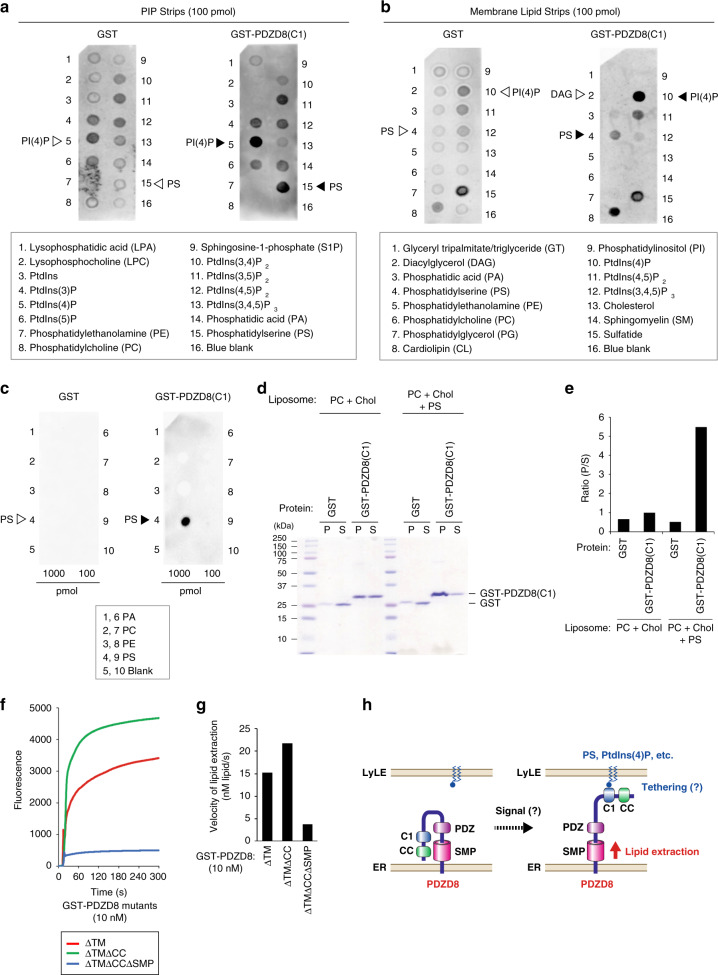
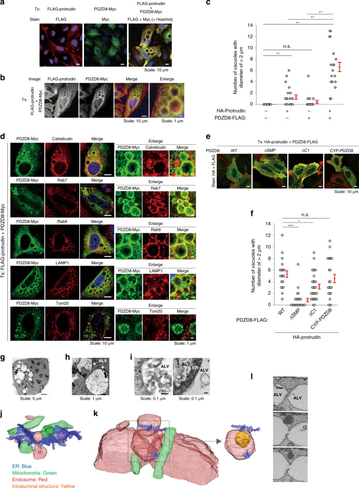
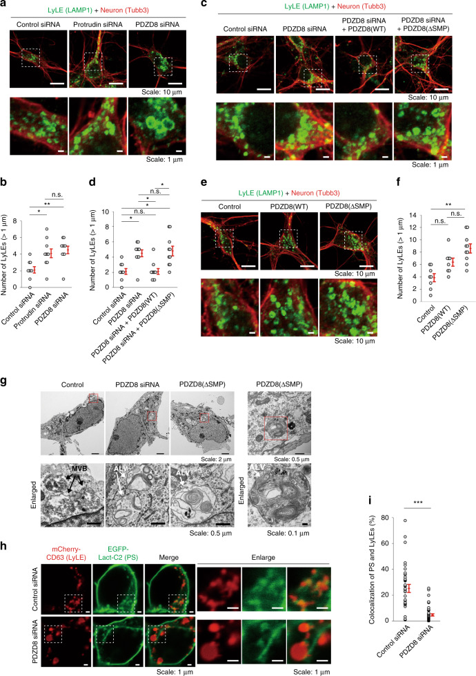
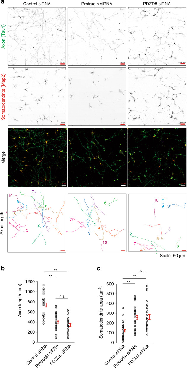
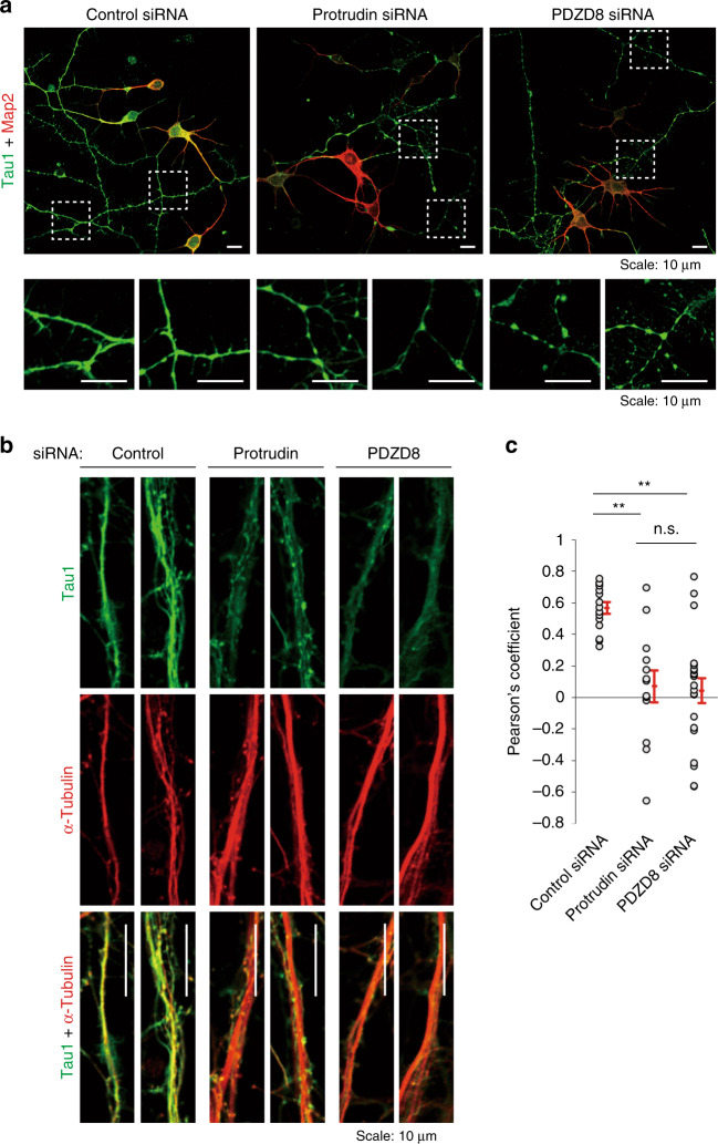
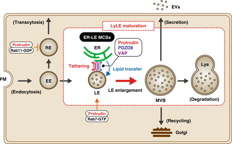
Endosome maturation depends on membrane contact sites (MCSs) formed between endoplasmic reticulum (ER) and endolysosomes (LyLEs). The mechanism underlying lipid supply for this process and its pathophysiological relevance remains unclear, however. Here, we identify PDZD8-the mammalian ortholog of a yeast ERMES subunit-as a protein that interacts with protrudin, which is located at ER-LyLE MCSs. Protrudin and PDZD8 promote the formation of ER-LyLE MCSs, and PDZD8 shows the ability to extract various lipids from the ER. Overexpression of both protrudin and PDZD8 in HeLa cells, as well as their depletion in mouse primary neurons, impairs endosomal homeostasis by inducing the formation of abnormal large vacuoles reminiscent of those apparent in spastin- or REEP1-deficient neurons. The protrudin-PDZD8 system is also essential for the establishment of neuronal polarity. Our results suggest that protrudin and PDZD8 cooperatively promote endosome maturation by mediating ER-LyLE tethering and lipid extraction at MCSs, thereby maintaining neuronal polarity and integrity.


Conflict of interest statement

The authors declare no competing interests.

Figures

Fig. 1
Fig. 1. Identification of protrudin-associated proteins with a proteomics approach.
a Extracts prepared from Neuro2A cells either stably expressing His6-FLAG-tagged mouse protrudin or syntaxin7 (negative control) or infected with the corresponding empty retrovirus (Vector) were subjected to dual affinity purification with antibodies to FLAG and Ni-NTA agarose. The purified proteins were separated by SDS-PAGE and stained with silver. Open and filled arrowheads indicate bands corresponding to bait proteins or protrudin binding proteins, respectively. b Extracts prepared from WT mouse brain were subjected to immunoprecipitation with antibodies to protrudin. The identified binding proteins were ranked according to the number of identified peptides from two independent experiments (Exp). c Extracts prepared from WT mouse brain were subjected to immunoprecipitation (IP) with antibodies to protrudin or to PDZD8 or with normal rabbit serum (NRS). The resulting precipitates were subjected to immunoblot (IB) analysis with antibodies to protrudin and to PDZD8. d, e Domain organization of human protrudin (d) and mouse PDZD8 (e), and structure of deletion mutants thereof. f Domain structure of mouse PDZD8 derivatives in which the TM domain is replaced with the corresponding domain of mouse CYP or Tom20, as well as of WT and ΔTM mutant forms of PDZD8. g, h Protein extracts prepared from the brain of WT (+/+) or PDZD8-deficient (−/−) mice (g), or from WT (+/+) or protrudin-deficient (−/−) mice (h), were subjected to immunoblot analysis with antibodies to protrudin, to PDZD8, and to calnexin (loading control). Filled and open arrowheads indicate bands corresponding to protrudin in PDZD8−/− mice and to PDZD8 in protrudin−/− mice, respectively. i PC12 cells transfected with protrudin, PDZD8, TMEM55B (negative control), or scrambled (Control) siRNAs were incubated in the absence or presence of 5 μM MG132 for 6 h. Immunoblot analysis was performed with antibodies to protrudin, to PDZD8, to TMEM55B, and to HSP90 (loading control). Filled and open arrowheads indicate blots corresponding to protrudin in cells transfected with PDZD8 siRNA or to PDZD8 in cells transfected with protrudin siRNA, respectively. The asterisks indicate nonspecific signals. We repeated all experiments at least three times independently with similar results.
Fig. 2
Fig. 2. Effects of protrudin and PDZD8 on tethering between the ER and LyLEs or mitochondria.
a Mouse primary cortical neurons cultured for 5 days in vitro were fixed, stained with antibodies to the ER marker PDI, to the LyLE marker LAMP1, and to PDZD8, and observed by super-resolution microscopy. Scale bars, 500 nm. b Rotated three-dimensional (3D) images derived from (a). Arrowheads in a and b indicate the localization of PDZD8 at ER-LyLE MCSs. c Manders overlap coefficient for colocalization of PDZD8 with either PDI (ER) or LAMP1 (LyLE) determined from images as in a. Data are means + SE for 14 cells. ***P < 0.001 (Student’s t-test). d Schematic representation of the mechanism of action of split-GFP (enclosed, left), and strategy for statistical analysis (right). A representative image is shown in Supplementary Fig. 8c. e, i HeLa cells stably expressing FLAG-tagged mouse protrudin or Myc-tagged mouse PDZD8, or those infected with the corresponding empty retrovirus (Vector), were transfected with expression vectors for split-GFP fragments—ERj1-GFP(1–10) and either LAMP1-GFP(11) (e) or Tom70-GFP(11) (i)—as well as for mCherry and were then imaged by confocal fluorescence microscopy. Scale bars, 10 µm. Enlarged images are also shown. Scale bars, 1 µm. f, j Split-GFP analysis for the ER and either LyLEs (data are means ± SE for 19 to 22 independent cells) (f) or mitochondria (Mito) (data are means ± SE for 44 to 52 independent cells) (j) of HeLa cells infected with retroviruses for protrudin, PDZD8, or both proteins. g, k Split-GFP analysis for the ER and either LyLEs (data are means ± SE for 18 independent cells) (g) or mitochondria (data are means ± SE for 16 to 18 independent cells) (k) of HeLa cells transfected with control, protrudin, or PDZD8 siRNAs. h Split-GFP analysis for the ER and LyLEs of HeLa cells infected with retroviruses for PDZD8(WT), PDZD8(ΔC1), or CYP-PDZD8 (data are means ± SE for 19 to 28 independent cells). *P < 0.05, **P < 0.01, ***P < 0.001; n.s., not significant (Kruskal–Wallis test followed by the Steel-Dwass multiple comparison test). The mRNA levels for endogenous human and exogenous mouse protrudin and PDZD8 are shown in Supplementary Fig. 9. We repeated all experiments at least three times independently with similar results.
Fig. 3
Fig. 3. Lipid extraction activity of PDZD8.
a Domain structure of mouse PDZD8(WT) and PDZD8(ΔTM). b Schematic representation of the liposome-FRET assay. c Phospholipid (PA, PS, PE, and PC) extraction activities of GST-PDZD8(ΔTM). d Ceramide (left) or cholesterol (right) extraction activity of GST-PDZD8(ΔTM). e Initial velocity of lipid extraction by GST-PDZD8(ΔTM) measured in c and d). f Phospholipid extraction activities of GST-PDZD8(ΔTM) with PA, PS, PE, and PC in the absence (−) or presence (+) of acceptor liposomes. g Concentration dependence for lipid extraction activity of GST-PDZD8(ΔTM) with PS in the absence (left) or presence (right) of acceptor liposomes. h Initial velocity of lipid extraction by GST-PDZD8(ΔTM) measured in g. i Detection of lipid (PS) extracted by GST-PDZD8(ΔTM) in the supernatent (Sup) separated from liposomes in the pellet after ultracentrifugation of the reaction mixture at 300 s.
Fig. 4
Fig. 4. Delineation of the region of PDZD8 responsible for lipid extraction.
a Domain structure of mouse PDZD8 deletion mutants. A summary of the lipid extraction activity of the mutants is shown on the right. PRT, protrudin; ND, not determined. b Phospholipid extraction activities of PDZD8 deletion mutants with PA, PS, PE, and PC. c Initial velocity of lipid extraction by PDZD8 mutants with PS measured in b. d Schematic representation of the liposome-FRET assay performed with DGS-NTA(Ni) and His6-tagged proteins. e PS extraction activity of His6-PDZD8(ΔTM) or His6-GST (negative control) measured with donor liposomes containing DGS-NTA(Ni) and in the absence (−) or presence (+) of acceptor (Acc) liposomes. f Initial velocity of lipid extraction by His6-PDZD8(ΔTM) measured in e.
Fig. 5
Fig. 5. Lipid-binding activity of PDZD8(C1) and mode of action of PDZD8.
ac Recombinant GST or GST-PDZD8(C1) was incubated with PIP Strips (a), Membrane Lipid Strips (b), or custom lipid strips (c), after which binding of the proteins to lipid spots on the strips was probed with antibodies to GST and immunoblot reagents. The lipids on the strips are listed below. d, e Recombinant GST or GST-PDZD8(C1) was incubated with liposomes consisting of PC and cholesterol (Chol), with or without PS, after which the liposomes were isolated by centrifugation and the resulting pellet (P) and supernatant (S) were subjected to SDS-PAGE followed by staining with Coomassie brilliant blue (d) and quantitative analysis of protein amount in each fraction (e). f Phospholipid extraction activity of the indicated PDZD8 deletion mutants with PS. g Initial velocity of lipid extraction by the mutants measured in f. h Model for the possible mode of action of PDZD8. PDZD8 is integrated into the ER membrane via its TM domain and tethers the LyLE membrane by binding to PS or PtdIns(4)P via its C1 domain. The CC domain inhibits lipid extraction activity of PDZD8. We repeated all experiments at least three times independently with similar results.
Fig. 6
Fig. 6. Abnormal large vacuole (ALV) formation induced by coexpression of protrudin and PDZD8.
a, b HeLa cells transiently transfected (Tx) with expression vectors for FLAG-protrudin, PDZD8-Myc, or both proteins were stained with antibodies to FLAG (red) and to Myc (green). Nuclei were also stained with Hoechst 33258 (blue). Scale bars, 10 or 1 µm as indicated. c Quantification of the number of vacuoles with a diameter of >2 µm in HeLa cells transiently transfected with expression vectors for HA-protrudin, PDZD8-FLAG, or both proteins. Data are means ± SE for 19 to 21 independent cells. **P < 0.01 (Kruskal–Wallis test followed by the Steel-Dwass multiple comparison test). d HeLa cells transiently transfected with expression vectors for FLAG-protrudin and PDZD8-Myc were stained with antibodies to Myc (green) as well as with those to the organelle marker proteins (red) calreticulin (ER), Rab7 or Rab9 (LEs), LAMP1 (LyLEs), or Tom20 (mitochondria). Merged images include nuclear staining with Hoechst 33258 (blue). Scale bars, 10 µm. The boxed regions are shown enlarged on the right. Scale bars, 1 µm. e, f HeLa cells transiently transfected with expression vectors for HA-protrudin as well as WT or the indicated mutant forms of PDZD8-FLAG were stained with antibodies to HA (red) and to FLAG (green) (e). Scale bar, 10 µm. The number of vacuoles with a diameter of >2 µm was quantitated (f). Data are means + SE for 20 to 29 independent cells. *P < 0.05, ***P < 0.001 (Kruskal–Wallis test followed by the Steel-Dwass multiple comparison test). gi HeLa cells transiently transfected with expression vectors for GFP-protrudin and PDZD8-GFP were imaged by TEM. Images of low (g), medium (h), or high (i) magnification are shown (i). Scale bars: 5 µm (g), 1 µm (h), or 0.1 µm (i). j, k Organelles of control cells (j) or of HeLa cells coexpressing FLAG-protrudin and PDZD8-Myc (k) were examined by FIB-SEM. The LE with an intraluminal structure in the boxed region of (k) is shown on the right. l Series of images along the z-axis used for FIB-SEM of HeLa cells coexpressing FLAG-protrudin and PDZD8-Myc. We repeated all experiments at least three times independently with similar results.
Fig. 7
Fig. 7. Effects of PDZD8 on LyLE maturation in neurons.
af Mouse primary cortical neurons transfected with control, protrudin, or PDZD8 siRNAs (a), with PDZD8 siRNA and expression vectors for siRNA-resistant forms of FLAG-tagged PDZD8(WT) or PDZD8(ΔSMP) (c), or with expression vectors for PDZD8(WT) or PDZD8(ΔSMP) (e) were cultured for 9 days in vitro, fixed, stained with antibodies to the neuronal marker Tubb3 and to the LyLE marker LAMP1, and observed with a confocal fluorescence microscope. Scale bars, 10 µm. The boxed regions in the upper panels are shown enlarged below. Scale bars, 1 µm. The number of LyLEs with a diameter of >1 µm in images as in (a), (c), and (e) are quantified in (b), (d), and (f), respectively. Data are means + SE for 8 to 10 independent cells. *P < 0.05, **P < 0.01 (Kruskal–Wallis test followed by the Steel-Dwass multiple comparison test). g Neurons transfected with PDZD8 siRNA or with an expression vector for PDZD8(ΔSMP) were cultured for 7 days in vitro, fixed, embedded in epoxy resin, and imaged by TEM. The boxed regions in the upper panels are shown at higher magnification in the lower panels. ALVs containing both ILVs and irregular intraluminal structures in cells expressing PDZD8(ΔSMP) are shown in the right panels. Scale bars are as indicated. h Neurons transfected with expression vectors for mCherry-CD63 and EGFP-Lact-C2, as well as with control or PDZD8 siRNAs were observed with a confocal fluorescence microscope after 6 days in vitro. (N = 44 to 52 for independent cells). Scale bars, 1 µm. The boxed regions in the left group of panels are shown enlarged on the right. Scale bars, 1 µm. i Quantification of the mCherry/EGFP colocalization ratio in images as in h. Data are means + SE for 36 to 53 independent regions. ***P < 0.001 (Student’s t-test). We repeated all experiments at least three times independently with similar results.
Fig. 8
Fig. 8. Effects of protrudin and PDZD8 on neuronal polarity.
a Mouse primary cortical neurons transfected with control, protrudin, or PDZD8 siRNAs were cultured in vitro for 5 days, fixed, stained with antibodies to the axonal marker Tau1 and to the somatodendrite marker Map2, and observed with a confocal fluorescence microscope. Axons were traced and are distinguished by number and color for measurement of their length (lowest panels). Asterisks indicate axons that extend beyond the area shown. Scale bars, 50 µm. b, c Axon length (b) and somatodendrite area (c) determined from images as in a with the use of the ImageJ-based program Fiji. Data are means + SE for 31 to 33 independent cells. *P < 0.05, **P < 0.01 (Kruskal–Wallis test followed by the Steel-Dwass multiple comparison test). We repeated all experiments at least three times independently with similar results.
Fig. 9
Fig. 9. Effects of protrudin and PDZD8 on neuronal integrity.
a Mouse primary cortical neurons transfected with control, protrudin, or PDZD8 siRNAs were cultured for 4 days in vitro, fixed, stained with antibodies to Tau1 and to Map2, and observed with a confocal fluorescence microscope. Scale bars, 10 µm. The boxed regions of the upper panels are shown enlarged below. Scale bars, 10 µm. b Neurons transfected as in a were cultured for 7 days in vitro, fixed, stained with antibodies to Tau1 and to α-tubulin, and observed with a confocal fluorescence microscope. Enlarged images of axons are shown. Scale bars, 10 µm. c Pearson’s colocalization coefficient for Tau1 and α-tubulin in images as in b. Data are means + SE for 13 to 21 independent regions. **P < 0.01 (Kruskal–Wallis test followed by the Steel-Dwass multiple comparison test). We repeated all experiments at least three times independently with similar results.
Fig. 10
Fig. 10. Protrudin and PDZD8 promote endosome maturation at ER-LyLE MCSs.
LEs undergo maturation to form MVBs through an increase in size and the formation of ILVs. This process is promoted by tethering of LyLEs to the ER via a VAP-protrudin- PDZD8 complex, with PDZD8 mediating a transfer of lipid from the ER to LyLEs. EVs, extracellular vesicles; Lys lysosome, PM plasma membrane, RE recycling endosome.

References

    1. Helle SC, et al. Organization and function of membrane contact sites. Biochim. Biophys. Acta. 2013;1833:2526–2541. - PubMed
    1. Phillips MJ, Voeltz GK. Structure and function of ER membrane contact sites with other organelles. Nat. Rev. Mol. Cell Biol. 2016;17:69–82. - PMC - PubMed
    1. Salvador-Gallego R, Hoyer MJ, Voeltz GK. SnapShot: functions of endoplasmic reticulum membrane contact sites. Cell. 2017;171:1224–1224. - PubMed
    1. Saheki Y, De Camilli P. Endoplasmic reticulum-plasma membrane contact sites. Annu. Rev. Biochem. 2017;86:659–684. - PubMed
    1. Wu, H., Carvalho, P. & Voeltz, G. K. Here, there, and everywhere: The importance of ER membrane contact sites. Science361, eaan5835 (2018). - PMC - PubMed

Publication types

MeSH terms