Case Reports
. 2021 May;478(5):977-984.
doi: 10.1007/s00428-020-02923-4. Epub 2020 Sep 12.

Primary glomus tumour of the pituitary gland: diagnostic challenges of a rare and potentially aggressive neoplasm

Affiliations
Case Reports

Primary glomus tumour of the pituitary gland: diagnostic challenges of a rare and potentially aggressive neoplasm

Boon Leong Quah et al. Virchows Arch. 2021 May.

Abstract

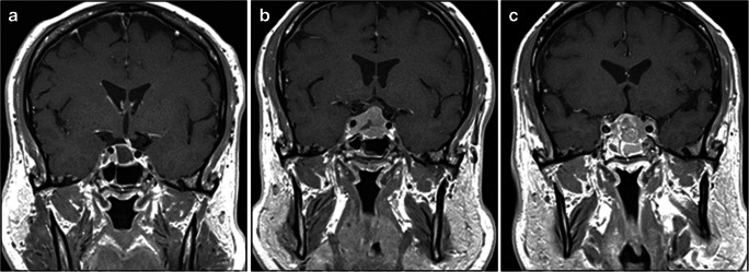
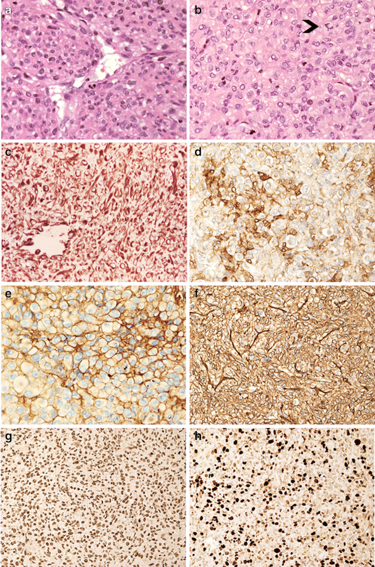
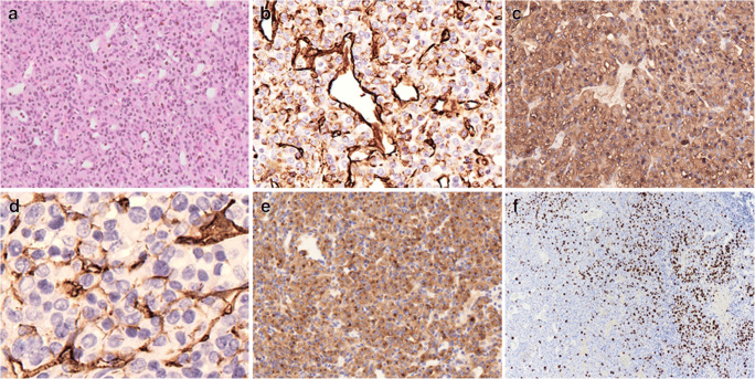
Primary non-neuroendocrine tumours of the pituitary gland and sella are rare lesions often challenging to diagnose. We describe two cases of clinically aggressive primary glomus tumour of the pituitary gland. The lesions occurred in a 63-year-old male and a 30-year-old female who presented with headache, blurred vision and hypopituitarism. Neuroimaging demonstrated large sellar and suprasellar tumours invading the surrounding structures. Histologically, the lesions were characterised by angiocentric sheets and nests of atypical cells that expressed vimentin, smooth muscle actin and CD34. Perivascular deposition of collagen IV was also a feature. Case 2 expressed synaptophysin. INI-1 (SMARCB1) expression was preserved. Both lesions were mitotically active and demonstrated a Ki-67 labelling index of 30%. Next-generation sequencing performed in case 1 showed no mutations in the reading frame of 37 commonly mutated oncogenes, including BRAF and KRAS. Four pituitary glomus tumours have previously been reported, none of which showed features of malignant glomus tumour. Similar to our two patients, three previous examples displayed aggressive behaviour.

Keywords: Glomus tumour; Malignant; Non-neuroendocrine tumour; Pituitary gland; Sella.


Conflict of interest statement

The authors declare no conflicts of interest.

Figures

Fig. 1
Fig. 1
(Patient 1) Preoperative coronal post-contrast T1 MRI documents spontaneous resolution of the sellar lesion with a residual largely sellar cystic lesion causing deviation of the pituitary stalk to the left. There is minimal bulging into the suprasellar cistern but no compromise on the optic chiasm (A). Coronal post-contrast T1 imaging after 4 months shows interval deterioration in appearances with a recurrent solid enhancing sellar mass invading the right cavernous sinus. There is also worsening extension into the suprasellar cistern with displacement of the optic chiasm superiorly (B). Post-contrast coronal MRI after surgery and radiotherapy shows a recurrent solid enhancing sellar mass invading the cavernous sinuses bilaterally and with suprasellar extension (C)
Fig. 2
Fig. 2
(Patient 2) MR imaging at presentation including (A) coronal T2, (B) post-contrast coronal T1 and (C) post-contrast sagittal T1 sequences documents a large heterogeneous mass lesion with heterogeneously enhancing solid and cystic components and intra-tumoural haemorrhage (A&B). The mass expands the sella and invades into the cavernous sinuses bilaterally. There is marked suprasellar extension and compression of the optic chiasm. The tumour bulges into the anterior aspect of the third ventricle (C) with secondary oedema and high T2 signal in the right frontal lobe (A)
Fig. 3
Fig. 3
(Patient 1) The lesion is composed of densely cellular sheets of tumour cells that form thin-walled vessels (A, HE, × 20). Neoplastic cells have epithelioid features and display eosinophilic cytoplasm and hyperchromatic nucleus; atypical mitoses are also present (arrowhead) (B, HE, × 40); vimentin is diffusely expressed (C, immunoperoxidase × 20); smooth muscle actin is focally expressed in tumour cells (D, immunoperoxidase, ×40); there is pericellular deposition of collagen IV (E, immunoperoxidase, × 40); tumour cells expressed CD34 (F, immunoperoxidase × 20) and retain INI-1 nuclear expression (G, immunoperoxidase × 20). About 30% of tumour cells express Ki-67 (H, immunoperoxidase, × 20)
Fig. 4
Fig. 4
(Patient 2) The tumour is composed of sheets of atypical cells in a network of thin-walled vessels (A, × 20 – HE); neoplastic cells show diffuse expression of vimentin (B, immunoperoxidase × 20) and smooth muscle actin (C, immunoperoxidase × 20), focal CD34 (D, immunoperoxidase × 40) and diffuse synaptophysin (E, immunoperoxidase × 20); increase in Ki-67 is focal with areas showing up to 30% of positive tumour cells (F, immunoperoxidase × 10)

References

    1. Hyrcza MD, Winer DA, Shago M, Au K, Zadeh G, Asa SL, Mete O. TFE3-expressing perivascular epithelioid cell neoplasm (PEComa) of the sella turcica. Endocr Pathol. 2017;28(1):22–26. doi: 10.1007/s12022-016-9434-7. - DOI - PubMed
    1. Cossu G, Brouland JP, La Rosa S, Camponovo C, Viaroli E, Daniel RT, Messerer M. Comprehensive evaluation of rare pituitary lesions: a single tertiary care pituitary center experience and review of the literature. Endocr Pathol. 2019;30(3):219–236. doi: 10.1007/s12022-019-09581-6. - DOI - PubMed
    1. Abushamat LA, Kerr JM, Lopes MBS, Kleinschmidt-DeMasters BK. Very unusual sellar/suprasellar region masses: a review. J Neuropathol Exp Neurol. 2019;78:673–684. doi: 10.1093/jnen/nlz044. - DOI - PubMed
    1. Thakkar K, Ramteke-Jadhav S, Kasaliwal R, Memon SS, Patil V, Thadani P, Lomte N, Sankhe S, Goel A, Epari S, Goel N, Lila A, Shah NS, Bandgar T. Sellar surprises: a single-centre experience of unusual sellar masses. Endocr Connect. 2020;9(2):111–121. doi: 10.1530/EC-19-0497. - DOI - PMC - PubMed
    1. Gombos Z, Zhang PJ. Glomus tumor. Arch Pathol Lab Med. 2008;132(9):1448–1452. doi: 10.1043/1543-2165(2008)132[1448:GT]2.0.CO;2. - DOI - PubMed

Publication types

MeSH terms

Substances